Review
. 2010 Aug;13(4):517-23.
doi: 10.1016/j.mib.2010.05.012. Epub 2010 Jun 17.

Viral hijacking of the host ubiquitin system to evade interferon responses

Affiliations
Review

Viral hijacking of the host ubiquitin system to evade interferon responses

Kasinath Viswanathan et al. Curr Opin Microbiol. 2010 Aug.

Abstract

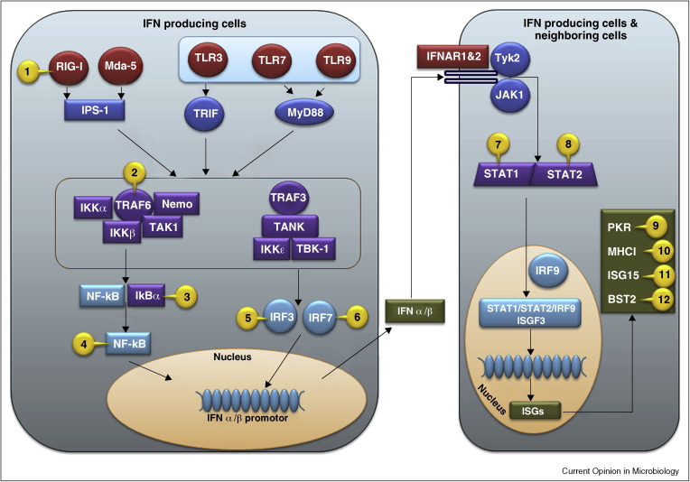
The post-translational attachment of ubiquitin or ubiquitin-like modifiers (ULMs) to proteins regulates many cellular processes including the generation of innate and adaptive immune responses to pathogens. Vice versa, pathogens counteract immune defense by inhibiting or redirecting the ubiquitination machinery of the host. A common immune evasion strategy is for viruses to target host immunoproteins for proteasomal or lysosomal degradation by employing viral or host ubiquitin ligases. By degrading key host adaptor and signaling molecules, viruses thus disable multiple immune response pathways including the production of and response to interferons as well as other innate host defense mechanisms. Recent work further revealed that viruses inhibit the ligation of ubiquitin or ULMs or remove ubiquitin from host cell proteins. Thus, viruses succeed in either stabilizing negative regulators of innate immune signaling or thwart host cell proteins that are activated by ubiquitin or ULM-modification.


Figures

Figure 1
Figure 1
Examples of viral protein mediated intervention of host pathogen stimulated IFN stimulation pathways using UPS. Host anti-viral signals are initiated when cells sense the presence of pathogens via pattern-recognition receptors (PRRs) such as extracellular toll like receptors (TLRs), or intracellular RIG-I like helicases (RLHs) or DNA binding proteins. PRRs recognize molecular signatures of pathogens, the single or double stranded RNA or DNA. TLR3 activates Toll–interleukin (IL)-1-resistance (TIR) domain-containing adaptor inducing IFN-b (TRIF), whereas TLR7 and 9 activate myeloid differentiation factor 88 (MyD88). Melanoma differentiation-associated gene (mda-5) and retinoic acid-inducible gene-1 (RIG-I) activate tumor necrosis factor (TNF) receptor associated factor 3 (TRAF3) via Cardif/VISA/MAVS/IPS-1. Activated TRAF3 interacts with TRAF family member associated NFκB activator (TANK), TANK binding kinase 1 (TBK-1), and the related IκB Kinase ɛ (IKKɛ) to phosphorylate and translocate IRFs in the nucleus. In a parallel pathway, NFκb activation is initiated by poly-ubiquitination of TRAF6 and receptor interacting protein-1 (RIP1). These adaptors recruit transforming growth factor (TGF) β—activating enzyme 1 (TAK1), NFκB essential modifier (NEMO), and IκB Kinases (IKK) into a multi protein complex that phosphorylates inhibitor of NFκB (IκB) resulting in its ubiquitination and degradation by proteasomes. The degradation of IκB reveals the nuclear localization signal of NFκB that then translocates to the nucleus. In the nucleus, activated IRFs and NFκB assemble at the IFNβ promoter together with ATF-2/c-jun and other co-factors to stimulate IFN transcription. IFNs are secreted and induce an anti-viral state in neighboring cells by binding to the receptors IFNAR 1 & 2 and activating the tyrosine kinases JAK1 and Tyk2 that in turn activate signal transducer and activator of transcription (STATs). Phosphorylated STAT1 and STAT2 heterodimerize and translocate to the nucleus, where they associate with IRF-9 to form the hetrotrimeric transcription factor ISGF3. Binding of ISGF3 to the IFN stimulated response element (ISRE) induces transcription of IFN stimulated genes (ISGs). Viral intervention of these pathway proteins are marked in the figure as numbers and listed below in the virus (protein)[ref] format: 1. Influenza A virus (NS1) [22••]; 2. Measles virus (P) [27]; 3. African swine fever virus (A238L) [25], Rotavirus (NSP1) [28]; 4.Murid herpesvirus-4 (ORF73) [26], Poxvirus (CP77) [23]; 5. classical swine fever virus [15], Bovine viral diarrhea virus [16], Rotavirus [18, 19], HIV-1 (Vpr, Vif) [20]; 6. Ebola virus (VP35) [21]; 7. Para influenza simian virus 5 [29, 30]; 8. Respiratory syncytial virus (NS1) [31]; 9. Hepatitis C virus [70]; 10. Human cytomegalovirus (US2, US11) [3], Kaposi's Sarcoma associated herpesvirus [KSHV] (K3, K5) [1, 2••]; 11. Influenza virus (NS1A) [42••, 46, 47, 48, 49], vaccinia virus (E3) [51], Coronavirus (PLP) [53]; 12. HIV-1 (Vpu) [57, 61, 64, 65, 66], KSHV (K5) [63].

Comment in

References

    1. Cadwell K., Coscoy L. Ubiquitination on nonlysine residues by a viral E3 ubiquitin ligase. Science. 2005;309:127–130. - PubMed
    1. Wang X., Herr R.A., Chua W.J., Lybarger L., Wiertz E.J., Hansen T.H. Ubiquitination of serine, threonine, or lysine residues on the cytoplasmic tail can induce ERAD of MHC-I by viral E3 ligase mK3. J Cell Biol. 2007;177:613–624. - PMC - PubMed
    2. References 1 and 2 demonstrate that non-lysine residues can be targeted for ubiquitination by E3 ligases.

    1. Isaacson M.K., Ploegh H.L. Ubiquitination, ubiquitin-like modifiers, and deubiquitination in viral infection. Cell Host Microbe. 2009;5:559–570. - PMC - PubMed
    1. Randow F., Lehner P.J. Viral avoidance and exploitation of the ubiquitin system. Nat Cell Biol. 2009;11:527–534. - PubMed
    1. Yu G.-Y., Lai M.M.C. The ubiquitin-proteasome system facilitates the transfer of murine coronavirus from endosome to cytoplasm during virus entry. J Virol. 2005;79:644–648. - PMC - PubMed