

VIP, CRF, and PACAP act at distinct receptors to elicit different cAMP/PKA dynamics in the neocortex
- PMID: 20699230
- PMCID: PMC3041014
- DOI: 10.1093/cercor/bhq143
VIP, CRF, and PACAP act at distinct receptors to elicit different cAMP/PKA dynamics in the neocortex
Abstract
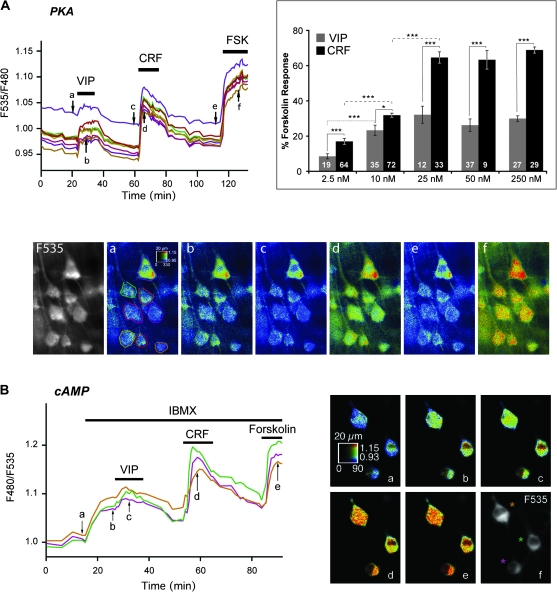
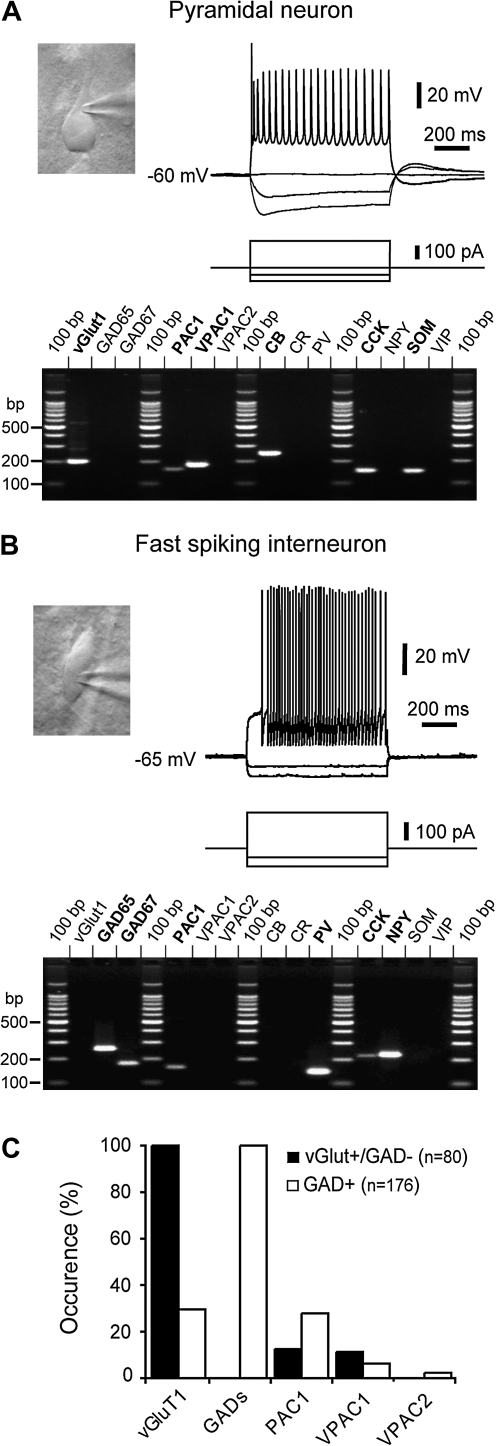
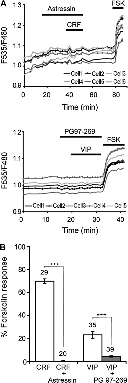
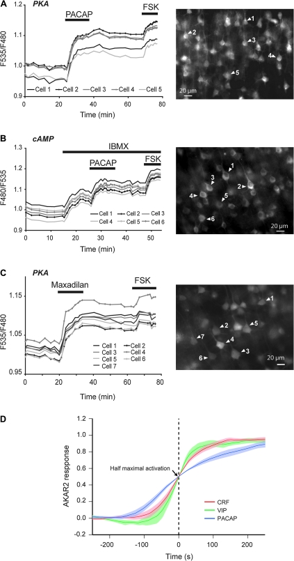
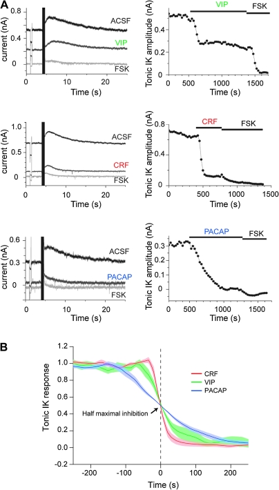
The functional significance of diverse neuropeptide coexpression and convergence onto common second messenger pathways remains unclear. To address this question, we characterized responses to corticotropin-releasing factor (CRF), pituitary adenylate cyclase-activating peptide (PACAP), and vasoactive intestinal peptide (VIP) in rat neocortical slices using optical recordings of cyclic adenosine monophosphate (cAMP) and protein kinase A (PKA) sensors, patch-clamp, and single-cell reverse transcription-polymerase chain reaction. Responses of pyramidal neurons to the 3 neuropeptides markedly differed in time-course and amplitude. Effects of these neuropeptides on the PKA-sensitive slow afterhyperpolarization current were consistent with those observed with cAMP/PKA sensors. CRF-1 receptors, primarily expressed in pyramidal cells, reportedly mediate the neocortical effects of CRF. PACAP and VIP activated distinct PAC1 and VPAC1 receptors, respectively. Indeed, a selective VPAC1 antagonist prevented VIP responses but had a minor effect on PACAP responses, which were mimicked by a specific PAC1 agonist. While PAC1 and VPAC1 were coexpressed in pyramidal cells, PAC1 expression was also frequently detected in interneurons, suggesting that PACAP has widespread effects on the neuronal network. Our results suggest that VIP and CRF, originating from interneurons, and PACAP, expressed mainly by pyramidal cells, finely tune the excitability and gene expression in the neocortical network via distinct cAMP/PKA-mediated effects.
Figures
Similar articles
-
VPAC2-R mediates the lipolytic effects of pituitary adenylate cyclase-activating polypeptide/vasoactive intestinal polypeptide in primary rat adipocytes.Endocrinology. 2005 Feb;146(2):744-50. doi: 10.1210/en.2004-0504. Epub 2004 Oct 28. Endocrinology. 2005. PMID: 15514088
-
Expression localisation and functional activity of pituitary adenylate cyclase-activating polypeptide, vasoactive intestinal polypeptide and their receptors in mouse ovary.Reproduction. 2007 Aug;134(2):281-92. doi: 10.1530/REP-07-0051. Reproduction. 2007. PMID: 17660238
-
Pituitary adenylate cyclase-activating polypeptide and PACAP receptor expression and function in the rat adrenal gland.Int J Mol Med. 2002 Mar;9(3):233-43. Int J Mol Med. 2002. PMID: 11836629
-
Pharmacology and functions of receptors for vasoactive intestinal peptide and pituitary adenylate cyclase-activating polypeptide: IUPHAR review 1.Br J Pharmacol. 2012 May;166(1):4-17. doi: 10.1111/j.1476-5381.2012.01871.x. Br J Pharmacol. 2012. PMID: 22289055 Free PMC article. Review.
-
[Physiological significance of pituitary adenylate cyclase-activating polypeptide (PACAP) in the nervous system].Yakugaku Zasshi. 2002 Dec;122(12):1109-21. doi: 10.1248/yakushi.122.1109. Yakugaku Zasshi. 2002. PMID: 12510388 Review. Japanese.
Cited by
-
Single Cell Multiplex Reverse Transcription Polymerase Chain Reaction After Patch-clamp.J Vis Exp. 2018 Jun 20;(136):57627. doi: 10.3791/57627. J Vis Exp. 2018. PMID: 29985318 Free PMC article.
-
Emerging pharmacological targets for alcohol use disorder.Alcohol. 2024 Dec;121:103-114. doi: 10.1016/j.alcohol.2024.07.007. Epub 2024 Jul 26. Alcohol. 2024. PMID: 39069210 Review.
-
Pituitary adenylate cyclase-activating polypeptide (PACAP) inhibits the slow afterhyperpolarizing current sIAHP in CA1 pyramidal neurons by activating multiple signaling pathways.Hippocampus. 2014 Jan;24(1):32-43. doi: 10.1002/hipo.22201. Epub 2013 Sep 30. Hippocampus. 2014. PMID: 23996525 Free PMC article.
-
Single-fluorophore biosensors based on conformation-sensitive GFP variants.FASEB J. 2014 Mar;28(3):1375-85. doi: 10.1096/fj.13-240507. Epub 2013 Dec 12. FASEB J. 2014. PMID: 24334549 Free PMC article.
-
Noradrenalin and dopamine receptors both control cAMP-PKA signaling throughout the cerebral cortex.Front Cell Neurosci. 2014 Aug 21;8:247. doi: 10.3389/fncel.2014.00247. eCollection 2014. Front Cell Neurosci. 2014. PMID: 25191229 Free PMC article.
References
-
- Baraban S, Tallent M. Interneuron diversity series: neuropeptides-endogenous regulators of neuronal excitability. Trends Neurosci. 2004;27:135–142. - PubMed
-
- Calebiro D, Nikolaev VO, Persani L, Lohse MJ. Signaling by internalized G-protein-coupled receptors. Trends Pharmacol Sci. 2010;31:221–228. - PubMed
Publication types
MeSH terms
Substances
Grants and funding
LinkOut - more resources
Full Text Sources
Molecular Biology Databases
