. 2010 Sep 1;123(Pt 17):2943-54.
doi: 10.1242/jcs.068817. Epub 2010 Aug 10.

CHMP2B mutants linked to frontotemporal dementia impair maturation of dendritic spines

Affiliations

CHMP2B mutants linked to frontotemporal dementia impair maturation of dendritic spines

Agnès Belly et al. J Cell Sci. .

Abstract

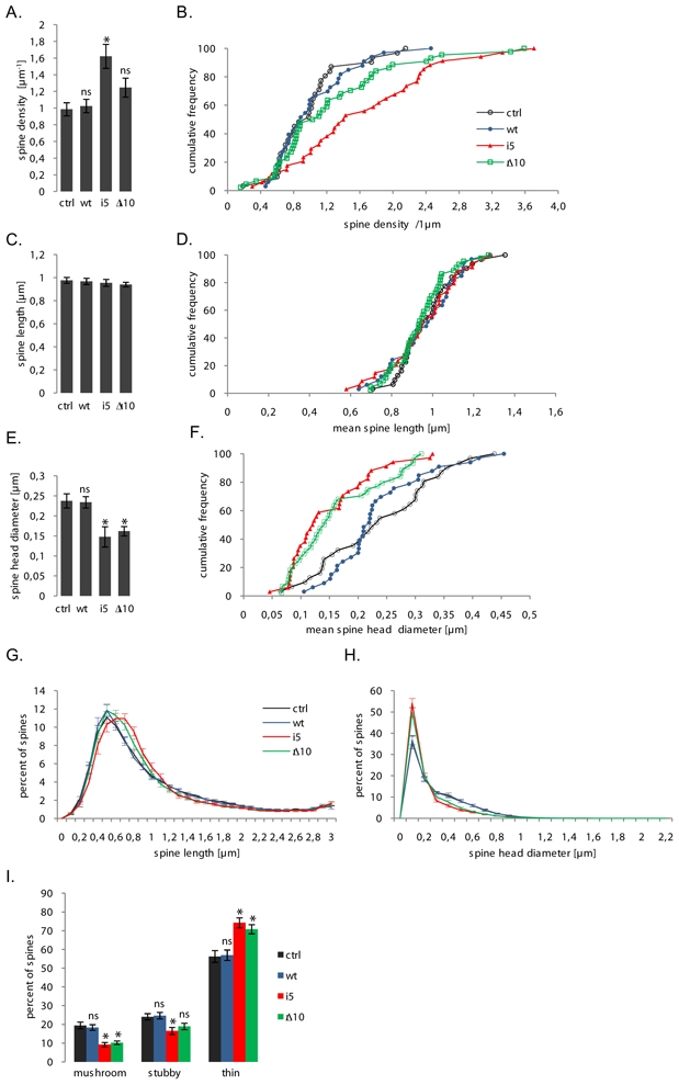
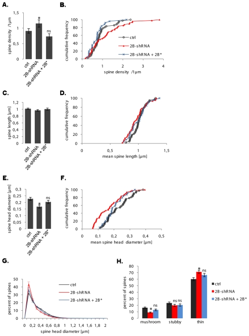
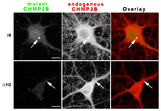
The highly conserved ESCRT-III complex is responsible for deformation and cleavage of membranes during endosomal trafficking and other cellular activities. In humans, dominant mutations in the ESCRT-III subunit CHMP2B cause frontotemporal dementia (FTD). The decade-long process leading to this cortical degeneration is not well understood. One possibility is that, akin to other neurodegenerative diseases, the pathogenic protein affects the integrity of dendritic spines and synapses before any neuronal death. Using confocal microscopy and 3D reconstruction, we examined whether expressing the FTD-linked mutants CHMP2B(intron5) and CHMP2B(Delta10) in cultured hippocampal neurons modified the number or structure of spines. Both mutants induced a significant decrease in the proportion of large spines with mushroom morphology, without overt degeneration. Furthermore, CHMP2B(Delta10) induced a drop in frequency and amplitude of spontaneous excitatory postsynaptic currents, suggesting that the more potent synapses were lost. These effects seemed unrelated to changes in autophagy. Depletion of endogenous CHMP2B by RNAi resulted in morphological changes similar to those induced by mutant CHMP2B, consistent with dominant-negative activity of pathogenic mutants. Thus, CHMP2B is required for spine growth. Taken together, these results demonstrate that a mutant ESCRT-III subunit linked to a human neurodegenerative disease can disrupt the normal pattern of spine development.


Figures

Fig. 1
Fig. 1
A: primary structure of CHMP2B constructs used in this study. The ESCRT-III domain is the conserved sequence domain common to all members of the Charged Multivesicular body Proteins (CHMP 1–7) superfamily. Positively (blue) and negatively (red) charged alpha helices (1 to 5) are located along the sequence according to the 3D structure of the closely related CHMP3 protein (Muziol et al., 2006). MIM: MIT (Microtubule Interacting and Transport domain) Interacting Module, required for binding to the SKD1/Vps4 ATPase responsible for dissociation of CHMP polymers. V: a single valine residue replaces the normal C terminus in CHMP2Bintron5. The green line in CHMP⊗10 indicates the aberrant C terminus carried by this mutant. Ellipses indicate tags fused to the N terminus (2×9 and 1×10 residues respectively). Sequence lengths are those of the untagged proteins. B: subcellular distribution of tagged CHMP2B proteins. Neurones were transfected with the indicated plasmids, processed for immunofluorescence staining with anti-tag antibody, and imaged by confocal microscopy. Two representative cells are shown for each plasmid. Top: CHMP2Bwt diffuses throughout the entire neuron, including the heads of dendritic spines (arrows). Middle: CHMP2Bintron5 forms multiple small clusters in the soma and dendrites, and occasional larger aggregates. Bottom: CHMP2BΔ10 has a clustered distribution similar to that of CHMP2Bintron5.
Fig. 2
Fig. 2
lack of LC3-GFP clustering in neurones expressing mutant CHMP2B. Neurones were co-transfected at 10 DIV with the indicated CHMP2B plasmids (left), together with the autophagosomal marker LC3-GFP. The cells were fixed 5 days later and processed for immunofluorescence staining with anti-tag antibody and Alexa594-coupled secondary antibody. Confocal images were acquired in both the immunofluorescence and GFP channels. Diffuse LC3-GFP localisation predominated upon cotransfection with CHMP2Bwt (a1,a2), CHMP2BΔ10 (c1,c2) and in a large majority of cases with CHMP2Bintron5 (b1,b2). Autophagosome accumulation was seen in rare cells transfected with CHMP2Bintron5 (b3, b4, arrow). Arrowhead: no autophagosomes were detected in the dendrites. Scale bars: 10 μm.
Fig. 3
Fig. 3
non-endosomal localisation of exogenously expressed CHMP2B proteins in hippocampal neurones. The neurones were doubly stained with monoclonal anti-tag antibody (a1– f1) together with polyclonal antibody against the respective marker (a2-f2), as indicated. Immunofluorescence was recorded by dual-channel confocal microscopy. Arrows indicate CHMP2B puncta. Note that they do not coincide with EEA1 (a2–c2) or LAMP1 (d2–f2) puncta. Scale bar: 10 μm.
Fig. 4
Fig. 4
detection and classification of dendritic spines in transfected hippocampal neurones. A: neurones were co-transfected with the indicated constructs, together with mCherry, fixed 5 days later, and images of mCherry fluorescence were acquired by confocal microscopy. The figure shows maximal intensity projections of confocal image stacks. Control: empty pcDNA3 vector. The boxed regions in a–d (scale bar: 10 μm) were scanned at higher magnification in a1–d1 (scale bar: 5 μm). B: stacks of optical slices acquired in A served to generate 3D models of the dendrite, using the surface-defining Ray Burst algorithm implemented in NeuronStudio (Rodriguez et al., 2006). The dimensions of the modelled structures were automatically measured as described by (Rodriguez et al., 2006), and used to identify and classify spines. Left: surface rendering of a segment from the dendrite shown in b1. Middle: maximal intensity projection of optical sections from the same segment. Right: automated spine detection and classification. Appropriately coloured dots are superimposed onto the spines. Yellow (t): thin spines; red(m): mushroom spines; blue (s): stubby spines.
Fig. 5
Fig. 5
effects of FTD-linked CHMP2B mutants on the density and dimensions of spines. All parameters were measured on the same sets of neurones (pooled from 3 independent cultures). Ctrl: control plasmid (n=31 neurones); wt: tagged wild-type CHMP2B (n=33 neurones); i5: CHMP2Bintron5 (n=34 neurones); Δ10: CHMP2BΔ10 (n=44 neurones). Measurements were obtained from 700–1500 μm dendrite and 600–1200 spines per neurone. A: mean spine counts per μm dendrite in neurones co-transfected with mCherry and the indicated plasmids. CHMP2Bintron5 increases the total density of spines (*p = 0.0005). The increase caused by CHMP2BΔ10 remains non-significant (ns) with p=0.09. B: cumulative frequency histogram of spine densities per neurone, corresponding to the dataset in A. C: mean length of spines per neurone. No significant difference was observed between conditions. D: cumulative frequency histogram of mean spine length per neuron, corresponding to dataset in C. E: Mean diameter of spine heads per neurone. Both mutants cause an overall shrinkage of spine heads (*p=0.0005 for both mutants). F: cumulative frequency histogram of mean spine head diameters per neuron, corresponding to the dataset in E. G: normalized distribution of spine lengths per neuron. Spine length histograms obtained for each neuron (0.1 μm-wide bins) were normalized to 100 total counts. The distribution shows the mean normalized values of each bin, for the respective neurone group. Error bars: S.E.M. for each bin. H: normalized distribution of spine head diameters per neurone. Spine diameter histograms obtained from each neuron were normalized and averaged as in F. Note the coincidence of the control and wild-type distributions, and that of the two mutant distributions. I: effects of FTD-linked CHMP2B mutants on the morphological type of spines. Histogram shows the mean percentages per neurone of spines with mushroom-like, stubby and thin morphologies. Both mutants potently reduce the mushroom spine fraction (*p<0.000001 for both mutants). CHMP2Bintron5 also reduces the fraction of stubby spines (*p=0.005) but CHMP2BΔ10 does not (p=0.09). Conversely, both mutants increase the thin spine fraction (*p=0.0001 for both mutants).
Fig. 6
Fig. 6
electrophysiological effects of wild-type and mutant CHMP2B. Miniature excitatory currents were recorded from transfected neurones under voltage clamp in the whole-cell configuration, in the presence of tetrodotoxin and D-APV to isolate AMPA receptor-mediated spontaneous synaptic currents (control: n=4 cells; CHMP2Bwt: n=6 cells; CHMP2BΔ10: n=6 cells). A: representative sample traces from neurones co-transfected with GFP and control plasmid (top), CHMP2Bwt (middle), and CHMP2BΔ10 (bottom). B: mean duration of inter-event intervals along sequences of mEPSCs pooled from equivalently transfected neurones (control: n=516 events; CHMP2Bwt: n=768 events; CHMP2BΔ10: n=768 events). Compared to control, intervals were significantly longer in mEPSCs from CHMP2BΔ10-expressing neurones (*p=0.00001), but not from CHMP2Bwt neurones (p=0.05, alpha=0.017). C: mean amplitude of mEPSCs from the different transfection groups. The overall variation of mean amplitude as a function of transfected plasmid was significant (ANOVA: F2,2049 = 3.04, p=0.048); the specific difference between the means of CHMP2BΔ10 and control was barely below threshold (*p=0.026 with Bonferroni-corrected alpha=0.025). D: cumulative histogram of mEPSC amplitudes; same dataset as in C. * indicates significant reduction in the top quintile in the case of CHMP2BΔ10 (see text and panel E). F: histogram of mEPSC amplitudes showing the specific reduction in the proportion of large currents (>20 pA) and the increase in small currents. S, i < 5pA; M, 5 <= i <=20 pA; L, i>20 pA. The two dashed lines refer to the respective proportions of large and small currents in the control neurones. The proportion of large currents was lower in neurones transfected with CHMP2BΔ10 (*p<0.05).
Fig. 7
Fig. 7
endogenous CHMP2B increases during neuronal maturation in culture. Sister cultures of hippocampal neurones maintained in plastic dishes were lysed at the indicated timepoints. The lysates were analysed by SDS-polyacrylamide gel electrophoresis and Western immunoblotting with anti-CHMP2B antibody. The blots were reprobed with anti-actin antibody to normalize for loading error. A: representative blot. Leftmost lane: rat brain lysate, showing the pattern of CHMP2B in vivo. B: films were scanned and the optical density of the CHMP2B band was measured in grey levels for each lane. Band intensities were normalized by dividing each value by the intensity of the actin band in the same lane. Care was taken to obtain immunodetection in the linear range. The graph shows mean normalized intensities at 3 timepoints (n=3 cultures). Error bar: S.E.M.
Fig. 8
Fig. 8
knock-down of endogenous CHMP2B in hippocampal neurones. A: to verify the efficacy of the shRNA-encoding plasmid, the indicated plasmids were transfected in rodent (BHK) cells and equal protein amounts of transfected cell lysates were analysed by Western immunoblotting with anti-CHMP2B antibody, or with anti-actin as a control. Lane 1: empty pSuper-mCherry plasmid; lane 2: shRNA-expressing plasmid; lane 3: shRNA-expressing plasmid cotransfected with vector encoding CHMP2B* (native CHMP2B cDNA with silent mutations at the siRNA target site). Note that the CHMP2B protein remaining after transfection of the shRNA plasmid largely originates from non-transfected cells in the culture. B: neurones were transfected at 10 DIV with the plasmids indicated on the left, fixed at 15 DIV, and stained with anti-CHMP2B antibody and Alexa488-labelled secondary antibody. Confocal images were acquired in both the Alexa488 and mCherry channels. Representative images of transfected neurones are displayed. Control: empty pSuper-mCherry vector. Note the drop in immunofluorescence in the shRNA-expressing neuron (arrow), compared to surrounding cells; and the decreased green vs. red ratio (overlay), compared to control or rescued cells. Scale bar: 10 μm. C: maximal intensity projections of image stacks, showing representative dendritic segments visualized by mCherry fluorescence in neurones transfected with the indicated plasmids.
Fig. 9
Fig. 9
effects of CHMP2B depletion on the density and morphology of dendritic spines. All parameters were measured as in Figs. 3 and 4. Ctrl: n=42 cells; shRNA: n=48 cells; shRNA together with rescuing CHMP2B: n=25 cells. A: the mean density of spines varied as a function of CHMP2B depletion (ANOVA: F 2,112 = 3.80, p = 0.025) although pairwise differences with control remained below significance (*p=0.021 vs. rescued cells). B: cumulative frequency histogram of spine densities per neurone (normalized in percent), same data as in A. C: CHMP2B depletion had no effect on the mean spine length per neuron. D: cumulative frequency histogram of spine lengths. E: mean spine head diameter per neurone. The CHMP2B shRNA induces a highly significant decrease (*p<0.001) which is rescued by re-expression of CHMP2B (ns, p=0.19). F: cumulative frequency histogram of spine head diameters. G: normalized distribution of spine head diameters. Note the superposition of “control” and “rescued” curves, and the recovery of larger spines in the “rescued” profile. H: proportions of basic spine types. The CHMP2B shRNA potently decreases the mushroom spine fraction (*p<0.001) and the effect is suppressed in the rescued cells (ns, p=0.028, corrected alpha = 0.017).
Fig. 10
Fig. 10
effect of mutant CHMP2B on the subcellular distribution of the normal protein. Hippocampal neurones were transfected with the indicated mutant, and processed for dual immunofluorescent staining with anti-tag and anti-endogenous CHMP2B antibodies. Confocal images were acquired in both channels. Co-clustering of endogenous CHMP2B with mutant protein (arrows) was evident in some of the CHMP2Bintron5 aggregates (upper panel), and faintly discernible in some of the CHMP2BΔ10 puncta (lower panel). Scale bars: 10 μm.

References

    1. Bajorek M, Schubert HL, McCullough J, Langelier C, Eckert DM, Stubblefield WM, Uter NT, Myszka DG, Hill CP, Sundquist WI. Structural basis for ESCRT-III protein autoinhibition. Nat Struct Mol Biol. 2009;16:754–62. - PMC - PubMed
    1. Banker G, Goslin K. MIT Press; 1998.
    1. Boyer C, Schikorski T, Stevens CF. Comparison of Hippocampal Dendritic Spines in Culture and in Brain. J Neurosci. 1998;18:5294–5300. - PMC - PubMed
    1. Carlton JG, Martin-Serrano J. Parallels between cytokinesis and retroviral budding: a role for the ESCRT machinery. Science. 2007;316:1908–12. - PubMed
    1. Fabrikant G, Lata S, Riches JD, Briggs JAG, Weissenhorn W, Kozlov MM. Computational Model of Membrane Fission Catalyzed by ESCRT-III. PLoS Comput Biol. 2009;5:e1000575. - PMC - PubMed

Publication types

Substances