. 2010 Sep 1;70(17):6804-14.
doi: 10.1158/0008-5472.CAN-10-0409. Epub 2010 Aug 10.

PIK3CA mutation uncouples tumor growth and cyclin D1 regulation from MEK/ERK and mutant KRAS signaling

Affiliations

PIK3CA mutation uncouples tumor growth and cyclin D1 regulation from MEK/ERK and mutant KRAS signaling

Ensar Halilovic et al. Cancer Res. .

Abstract

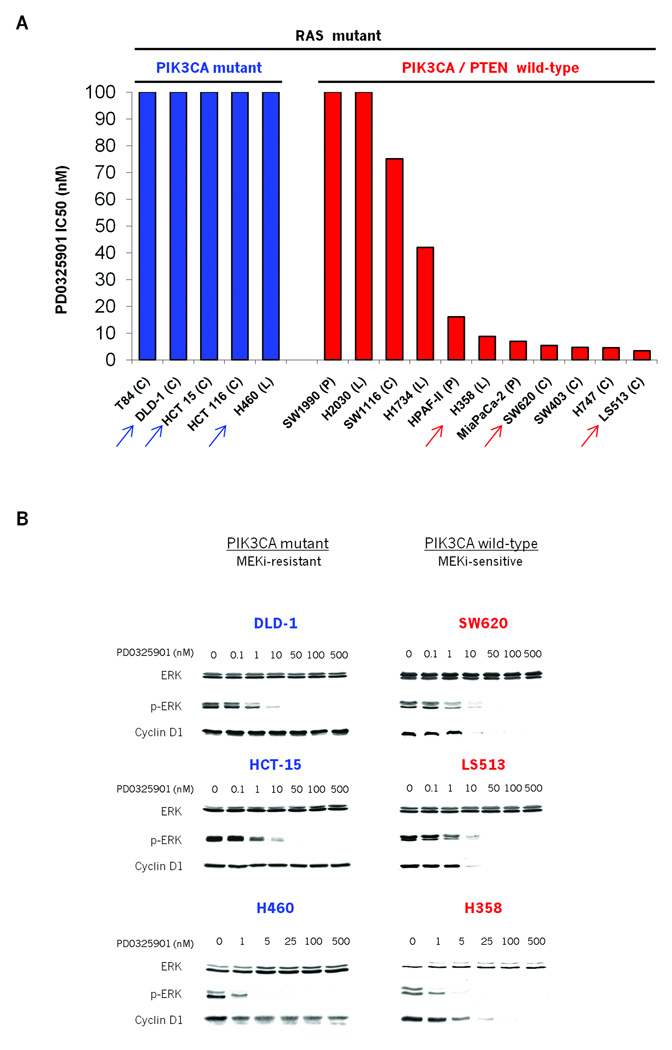
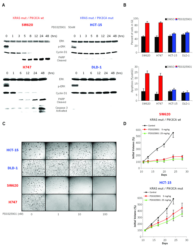
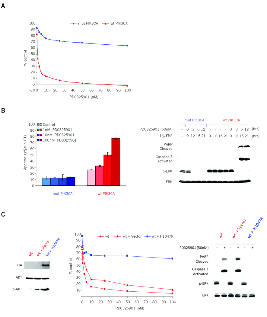
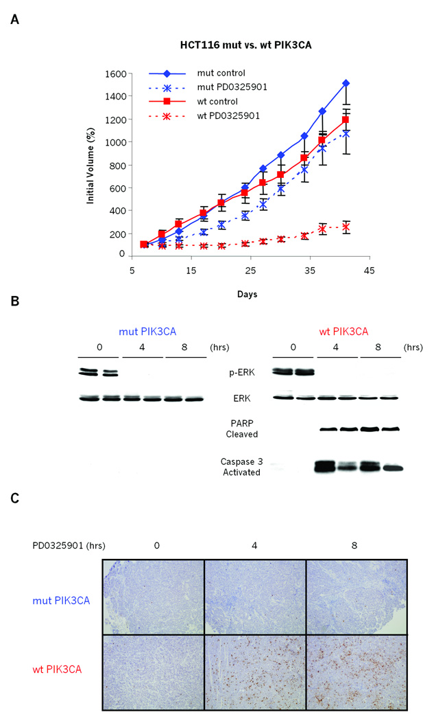
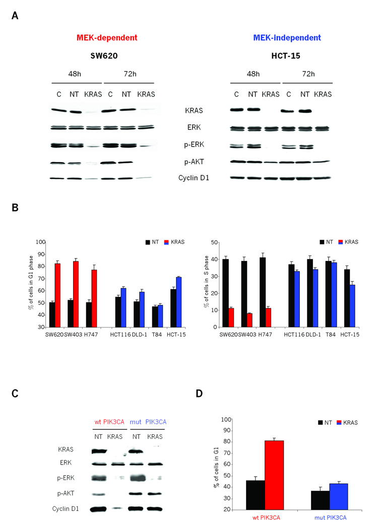
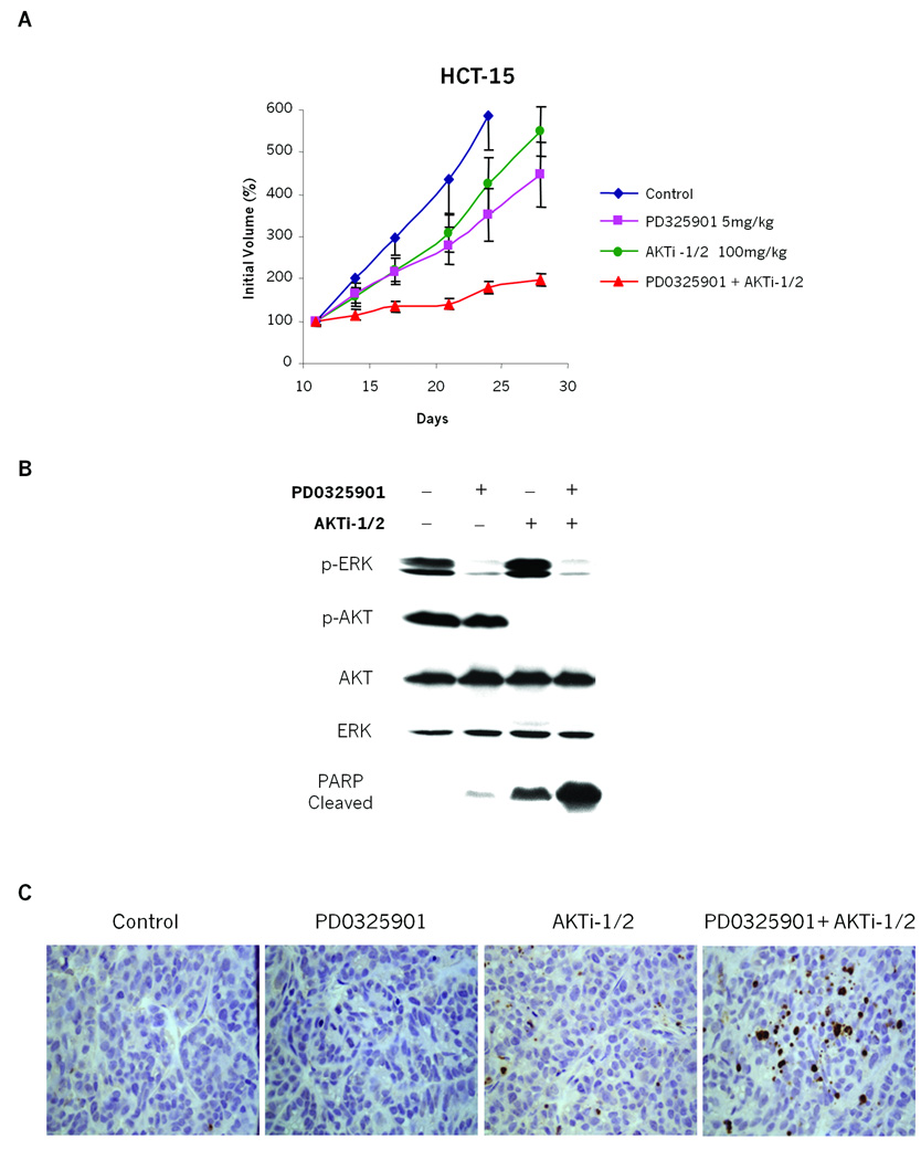
Mutational activation of KRAS is a common event in human tumors. Identification of the key signaling pathways downstream of mutant KRAS is essential for our understanding of how to pharmacologically target these cancers in patients. We show that PD0325901, a small-molecule MEK inhibitor, decreases MEK/ERK pathway signaling and destabilizes cyclin D1, resulting in significant anticancer activity in a subset of KRAS mutant tumors in vitro and in vivo. Mutational activation of PIK3CA, which commonly co-occurs with KRAS mutation, provides resistance to MEK inhibition through reactivation of AKT signaling. Genetic ablation of the mutant PIK3CA allele in MEK inhibitor-resistant cells restores MEK pathway sensitivity, and re-expression of mutant PIK3CA reinstates the resistance, highlighting the importance of this mutation in resistance to therapy in human cancers. In KRAS mutant tumors, PIK3CA mutation restores cyclin D1 expression and G(1)-S cell cycle progression so that they are no longer dependent on KRAS and MEK/ERK signaling. Furthermore, the growth of KRAS mutant tumors with coexistent PIK3CA mutations in vivo is profoundly inhibited with combined pharmacologic inhibition of MEK and AKT. These data suggest that tumors with both KRAS and phosphoinositide 3-kinase mutations are unlikely to respond to the inhibition of the MEK pathway alone but will require effective inhibition of both MEK and phosphoinositide 3-kinase/AKT pathway signaling.


Figures

Figure 1
Figure 1. MEK/ERK dependence of tumor cell lines with mutant KRAS
A: Colon (C), pancreatic (P) and lung cancer (L) cell lines harboring KRAS mutation were grown in the presence of the MEK inhibitor PD0325901. Day 5 IC50 values were determined using Alamar Blue. Arrows indicate the representative cell lines illustrated in Fig 1B. B: Effects of PD0325901 on ERK, p-ERK and Cyclin D1 expression in MEK-inhibitor sensitive (red) and resistant (blue) cell lines. Cells were treated with PD0325901, harvested 24hrs post treatment and cell lysates immunoblotted with the indicated antibodies.
Figure 2
Figure 2. Coexistent PIK3CA mutation is associated with decreased dependency on MEK signaling for tumor growth in vitro and in vivo
A: Immunoblots showing the effects of MEK inhibition in PD0325901 sensitive (SW620, H747) and resistant (HCT-15, DLD-1) cell lines. Cells were treated with 50nM PD0325901 and lysates immunoblotted with the specified antibodies. B: MEK-dependent (SW620, H747) and MEK-independent (HCT-15, DLD-1) cells were treated with 50nM PD0325901 or DMSO control and harvested 48 hours later. Graphs show: (Top) the percent of cells with G1 DNA content and (Bottom) the apoptotic fraction reported as the percent of cells with sub-G1 DNA content. The results represent the mean ± SE of two independent experiments done in triplicate. C: MEK-dependent (SW620, H747) and MEK-independent (HCT-15, DLD-1) cells were treated with PD0325901 in a soft agar colony formation assay. Representative images at 21 days post plating are shown. D: Mice with established SW620 and HCT15 xenografts were treated with PD0325901 × 5 days/week, for 3 weeks, or with vehicle only. The results represent the mean percent increase in tumor volume ± SE (n = 5 mice/group).
Figure 3
Figure 3. Coexistent PIK3CA mutation causes KRAS mutant colorectal tumor cells to grow and survive in a MEK/ERK independent manner
A: Isogenic HCT116 KRAS mutant/PIK3CA wild-type (wt PIK3CA, red) and HCT116 KRAS mutant/PIK3CA mutant (mut PIK3CA, blue) cells were grown in the presence of various concentrations of PD0325901 in 1% FBS growth medium and proliferation was measured using Alamar Blue. Results are represented as percent of untreated control on day 3 plotted as a function of the drug concentration. B: Left panel: Apoptotic fraction of PD0325901 treated PIK3CA isogenic HCT116 cells, reported as the percent of cells with sub-G1 DNA content 48 hr post treatment. The results represent the mean ± SE of two independent experiments performed in triplicate. Right panel: PIK3CA isogenic HCT116 cells were grown in 1% FBS growth media for 9 hours and then treated with 50nM PD0325901. The cells were harvested at the indicated times post treatment, lysed and immunoblotted with the specified antibodies. C: Left panel: Immunoblots of HCT116 KRAS mutant/PIK3CA wild-type cells (wt, red) transduced with H1047R-mutant PIK3CA cDNA (wt+H1047R, blue) or vector-only control (wt+Vector, red). The stable expression was confirmed by immunoprecipitation and western blot with an anti-HA tag antibody. Middle panel: HCT116 KRAS mutant/PIK3CA wild-type cells (wt, red) and cells stably expressing mutant PIK3CA (wt + H1047R, blue) or vector-only (wt + vector, red) were grown in the presence of various concentrations of PD0325901 in 1% FBS growth medium and proliferation was measured using Alamar Blue. Results are represented as the percentage of untreated control on day 3, plotted as a function of the drug concentration. Right panel: Western blot of apoptotic markers and p-ERK in HCT116 wild-type PIK3CA cells (wt) and these cells with either stably transduced mutant PIK3CA (H1047R) or control vector in the presence of PD0325901. The cells were grown in 1% FBS growth media for 9 hours and then treated with 50nM PD0325901. The cells were harvested 12 hours post treatment, lysed and immunoblotted with the specified antibodies.
Figure 4
Figure 4. PIK3CA mutation causes mutant KRAS colorectal tumor HCT116 to grow in vivo in a MEK-independent manner
A: Mice with established HCT116 KRAS mutant/PIK3CA wild-type (wt PIK3CA, red) and HCT116 KRAS mutant/PIK3CA mutant (mut PIK3CA, blue) xenografts were treated with 5mg/kg of PD0325901 or vehicle only, 5 days/week for the indicated number of days. The results are represented as in Figure 2D. B and C: Immunoblots of homogenized xenograft tissue after a single 5mg/kg dose of PD0325901. Tumors were excised pretreatment and at indicated times and split in half; (B) one half was flash frozen for immunoblot analysis with indicated antibodies; (C) the other half was formalin fixed, paraffin embedded and used for tissue-section preparation. 8 µm tissue sections were cut and stained with human specific antibody for cleaved PARP.
Figure 5
Figure 5. MEK-dependent Cyclin D1 expression is also dependent on expression of KRAS in cells with wild-type PIK3CA
A: Immunoblots showing the effects of KRAS siRNA in both MEK-dependent (SW620) and MEK-independent (HCT15) cells. Cells were transfected with lipid carrier control (C), non-targeting control (NT) or KRAS siRNA and harvested at 48 and 72 hours post-transfection. Cell lysates were immunoblotted with the indicated antibodies. B: MEK-dependent (SW620, SW403, H747) and MEK-independent (HCT116, DLD-1, T-84, HCT-15) cells were transfected with either non-targeting (NT) or KRAS siRNA, and collected 48hr later. Nuclei were isolated, stained with ethidium bromide, and cell cycle distribution analyzed by flow cytometry. Graphs show: 1) the percent of cells with G1 DNA content and 2) the percent of cells with S phase DNA content. The results represent the mean ± SE of two independent experiments performed in triplicate. C and D: PIK3CA wild-type and mutant isogenic HCT116 cells were transfected with non-targeting control (NT) or KRAS siRNA, harvested 48 hr post transfection and (C) cell lysates immunoblotted with the indicated antibodies or (D) analyzed by flow cytometry. The results are represented as in Figure 5B.
Figure 6
Figure 6. Combined inhibition of both MEK/ERK and PI3K/AKT pathways shows benefits in suppressing growth of tumors with coexisting KRAS and PIK3CA mutations
A: Mice with established HCT15 (KRAS mutant/PIK3CA mutant) xenografts were dosed with 5mg/kg of PD0325901 and 100mg/kg of AKTi-1/2 alone or in combination. The results are represented as in Figure 2D. B and C:Immunoblots of homogenized xenograft tissue. Tumors were excised 8 hours post treatment and split in half; (B) one half was flash frozen for immunoblot analysis with indicated antibodies; (C) the other half was formalin fixed, paraffin embedded and used for analysis by TUNEL assay (described in Gavrieli et al. (32)).

References

    1. Shields JM, Pruitt K, McFall A, Shaub A, Der CJ. Understanding Ras: 'it ain't over 'til it's over'. Trends Cell Biol. 2000;10:147–154. - PubMed
    1. Campbell PM, Der CJ. Oncogenic Ras and its role in tumor cell invasion and metastasis. Semin Cancer Biol. 2004;14:105–114. - PubMed
    1. Bowen DT, Frew ME, Hills R, et al. RAS mutation in acute myeloid leukemia is associated with distinct cytogenetic subgroups but does not influence outcome in patients younger than 60 years. Blood. 2005;106:2113–2119. - PubMed
    1. Repasky GA, Chenette EJ, Der CJ. Renewing the conspiracy theory debate: does Raf function alone to mediate Ras oncogenesis? Trends Cell Biol. 2004;14:639–647. - PubMed
    1. Moodie SA, Willumsen BM, Weber MJ, Wolfman A. Complexes of Ras.GTP with Raf-1 and mitogen-activated protein kinase kinase. Science. 1993;260:1658–1661. - PubMed

Publication types

MeSH terms