. 2010 Nov;18(11):1917-26.
doi: 10.1038/mt.2010.167. Epub 2010 Aug 10.

POD nanoparticles expressing GDNF provide structural and functional rescue of light-induced retinal degeneration in an adult mouse

Affiliations

POD nanoparticles expressing GDNF provide structural and functional rescue of light-induced retinal degeneration in an adult mouse

Sarah P Read et al. Mol Ther. 2010 Nov.

Abstract

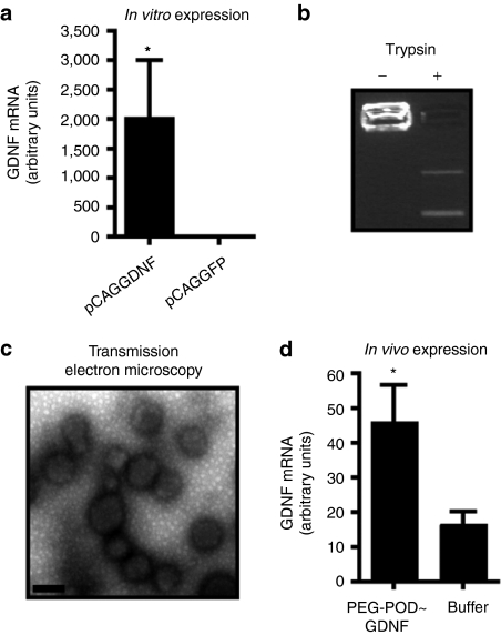
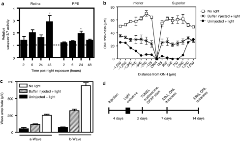
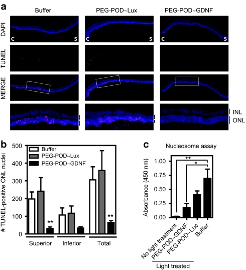
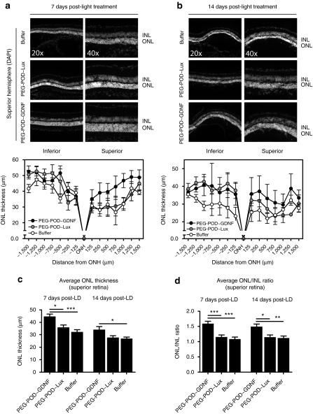
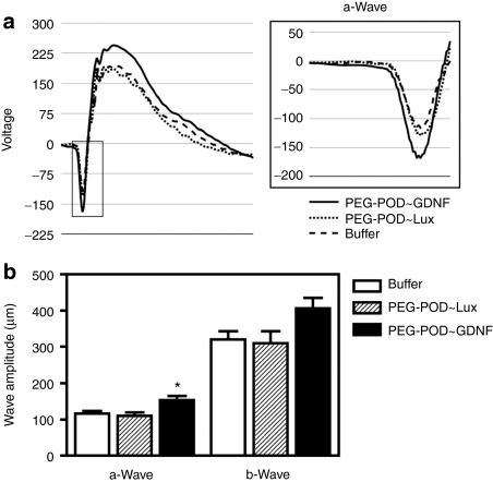
Peptide for ocular delivery (POD) is a novel cationic cell-penetrating peptide (CPP) which, when conjugated with polyethylene glycol (PEG-POD), can deliver plasmid DNA to the retinal pigment epithelium (RPE) of adult murine retina. PEG-POD nanoparticles containing an expression cassette for glial cell line-derived neurotrophic factor (PEG-POD~GDNF) were investigated for their ability to inhibit light-induced photoreceptor apoptosis. PEG-POD~GDNF, control nanoparticles, or buffer were injected into the subretinal space of adult murine retina and retinal degeneration induced by blue light. Animals injected with PEG-POD~GDNF showed a significant reduction (3.9-7.7 fold) in apoptosis relative to control-injected animals. The thickness of the outer nuclear layer (ONL) of the superior retina of PEG-POD~GDNF-injected eyes was significantly greater (23.6-39.3%) than control-injected retina 14 days post-light treatment. PEG-POD~GDNF-injected eyes showed a 27-39% greater functional response relative to controls, as measured by electroretinogram (ERG) 7 days post-light treatment. This is one of only two studies demonstrating histological and functional rescue of a mouse model of retinal degeneration following nonviral administration of a transgene into adult retina. Although rescue is short lived for clinical application, this study represents an important step in the development of nonviral gene therapy for retinal diseases.


Figures

Figure 1
Figure 1
PEG-POD forms GDNF-expressing nanoparticles. (a) A plasmid (pCAGGDNF) containing an expression cassette for rat GDNF was shown to express GDNF mRNA in vitro. Both conditions, n = 3. Mean ± SEM. (b) PEG-POD compaction of pCAGGDNF prevents electrophoretic migration of the plasmid, which can be relieved by trypsin-mediated digestion of the protein. (c) PEG-POD compacted pCAGGDNF (PEG-POD~GDNF) was examined by transmission electron microscopy (TEM) and found to form discrete spherical particles, similar to those previously described. Analysis of the TEM images showed a mean particle diameter of 175.9 ± 28.6 nm. Bar = 200 nm. (d) Injection of PEG-POD compacted pCAGGDNF into the subretinal space results in a detectable level of rat GDNF mRNA expression (P < 0.05). Both conditions, n = 9. Mean ± SEM. GDNF, glial cell line–derived neurotrophic factor; PEG, polyethylene glycol; POD, peptide for ocular delivery.
Figure 2
Figure 2
Bright blue light induces caspase-3/7 activation and retinal degeneration that is modulated by subretinal injection. (a) Caspase-3/7 activity was measured at various time points following light treatment. There was a 2.9-fold increase in activity in the retina 48 hours post-light exposure (P < 0.05), as well as a 1.9-fold increase in activity in the RPE 24 hours post-light exposure (P < 0.05), relative to the respective tissues of nonlight-treated mice. Two and forty-eight hours, n = 4; 6 and 24 hours, n = 6. Mean ± SEM. (b) Photoreceptor degeneration was evaluated by measuring the thickness of the ONL. The ONL of uninjected mice showed marked loss of photoreceptor nuclei post-light treatment, which was partially rescued by a subretinal injection of buffer. (c) Electroretinograms (ERGs) were recorded in mice 7 days after light exposure. In the absence of any injection, there was a significant decrease in the amplitudes of both the a-wave (P < 0.001) and the b-wave (P < 0.001). The impairment in light-response was partially prevented by subretinal injection of a buffer solution, although both the a- and b-waves were significantly lower in amplitude than those of nonlight-treated mice (P < 0.001). Buffer, n = 11; uninjected, n = 3; nonlight-treated, n = 4. Mean ± SEM. (d) To examine the effects of PEG-POD~GDNF, retinal degeneration was induced 4 days following subretinal injection by exposure to 4 hours of bright blue light. Mice were injected into the superior hemisphere with either PEG-POD~GDNF nanoparticles, PEG-POD~Lux (sham nanoparticles), or buffer alone. Following light exposure, effects of the treatment were assessed by measuring Müller cell activation (GFAP), apoptosis (TUNEL and nucleosome release), and ONL thickness and functional response (ERG) at 2, 7, and 14 days post-light-treatment as indicated. GFAP, glial fibrillary acidic protein; ONH, optic nerve head; ONL, outer nuclear layer; TUNEL, TdT-dUTP terminal nick-end labeling.
Figure 3
Figure 3
PEG-POD~GDNF nanoparticles result in decreased apoptosis of photoreceptors. (a) TUNEL stain of retinal sections 48 hours after light exposure showed a significant reduction in apoptotic photoreceptors in PEG-POD~GDNF-injected eyes. C, central; S, superior. (b) TUNEL-positive nuclei in the ONL of both the superior and inferior hemispheres. Eyes injected with PEG-POD~GDNF showed a significant decrease in the number of TUNEL-positive nuclei in the ONL of the superior pole (31.00 ± 8.5 nuclei/section) compared to PEG-POD~Lux (241.2 ± 77.6 nuclei/section, P < 0.01) and buffer- (197.9 ± 39.7, P < 0.01) injected eyes. PEG-POD~GDNF, n = 10; PEG-POD~Lux, n = 5; buffer, n = 7. Mean ± SEM. (c) Retinal tissue was harvested 48 hours post-light treatment and apoptosis evaluated by nucleosome release ELISA. Lower absorbance is observed with less nucleosome release and is thus an indicator of decreased apoptosis. Eyes injected with PEG-POD~GDNF showed 2.3-fold lower absorbance than PEG-POD~Lux-injected eyes (P > 0.05) and 3.9-fold lower than buffer-injected eyes (P < 0.05). Nonlight treated and PEG-POD~GDNF, n = 5; PEG-POD~Lux, buffer, and no light treatment, n = 5. Mean ± SEM. GDNF, glial cell line–derived neurotrophic factor; INL/ONL, inner/outer nuclear layer; PEG, polyethylene glycol; POD, peptide for ocular delivery; TUNEL, TdT-dUTP terminal nick-end labeling.
Figure 4
Figure 4
Injection of PEG-POD~GDNF results in decreased photoreceptor cell loss. To examine the effect on photoreceptor cell loss, ONL thickness was measured in retinas injected with either PEG-POD~GDNF, PEG-POD~Lux, or buffer harvested (a) 7 days or (b) 14 days post-light exposure. ONL thickness was measured at 250 µm intervals extending from the optic nerve. Representative images of the superior hemisphere adjacent to the ONH are shown. Average thickness of the ONL of the superior retina was calculated from measurements taken between 250 and 1,500 µm from the optic nerve. Similar measurements were taken for the INL and the average ONL/INL ratio calculated. (c) PEG-POD~GDNF-injected eyes showed a significant increase in the ONL thickness of the superior hemisphere by 24.5% when compared to PEG-POD~Lux (P < 0.05) or by 39.3% buffer (P < 0.001)-injected eyes 7 days post-light treatment and 27.7% relative to buffer (P < 0.05) 14 days post-light treatment. (d) Similar results were obtained when analyzing the ONL/INL ratio of the superior hemisphere. At 7 days the ratio was higher in eyes injected with PEG-POD~GDNF by 38.5% compared to those injected with PEG-POD~Lux (P < 0.001) or 47.3% compared to buffer (P < 0.001). The ONL/INL ratio remained higher at 14 days post-light exposure in PEG-POD~GDNF-treated eyes by 30.4% compared to PEG-POD~Lux (P < 0.05) and by 33.9% compared to buffer (P < 0.01). 7 days: PEG-POD~GDNF and PEG-POD~Lux, n = 6; buffer, n = 4. 14 days: PEG-POD~GDNF, n = 4; PEG-POD~Lux, n = 7; buffer, n = 6. Mean ± SEM. DAPI, 4′,6-diamidino-2-phenylindole; GDNF, glial cell line–derived neurotrophic factor; INL, inner nuclear layer; LD, light degeneration; ONH, optic nerve head; ONL, outer nuclear layer; PEG, polyethylene glycol; POD, peptide for ocular delivery.
Figure 5
Figure 5
Injection of PEG-POD~GDNF results in partial functional rescue of light-damaged retina. In order to evaluate the functional rescue of PEG-POD~GDNF-treated retinas, electroretinograms (ERGs) were performed 7 days post-light exposure. (a) Averages of scotopic ERG recordings for all eyes in each treatment condition. The a-wave is boxed in gray and shown in more detail. (b) The amplitude of both the a- and b-waves were measured and compared between injection conditions. PEG-POD~GDNF-injected eyes showed a 39% increase in amplitude in the a-wave compared to PEG-POD~Lux (P < 0.05) and 32% compared to buffer injected eyes (P < 0.01). Though not significant, the mean b-wave amplitude was increased in PEG-POD~GDNF-injected eyes by 31% compared to PEG-POD~Lux and by 27% compared to buffer-injected eyes. PEG-POD~GDNF, n = 12; PEG-POD~Lux, n = 9; buffer, n = 11. Mean ± SEM. GDNF, glial cell line-derived neurotrophic factor; PEG, polyethylene glycol; POD, peptide for ocular delivery.
Figure 6
Figure 6
Injection of PEG-POD~GDNF nanoparticles causes Müller cell activation. (a) Sections of retinas injected with PEG-POD~GDNF, PEG-POD~Lux, or buffer were stained for GFAP and compared to GFAP staining of retinas that had not been exposed to light. Eyes treated with light showed a redistribution of GFAP (arrows) regardless of injection condition, whereas nonlight-treated eyes showed a restriction of GFAP localization to astrocytes and the end feet of Müller cells at the inner limiting membrane. Eyes injected with PEG-POD~GDNF showed an increased level of GFAP staining throughout the Müller cell extending toward the outer limiting membrane (arrows) compared to other injections. (b) Quantification of GFAP fluorescence in the inner retina revealed a significant increase in GFAP in the inner retina in PEG-POD~GDNF injected eyes compared to both injection controls (P < 0.05). PEG-POD~GDNF, n = 7; PEG-POD~Lux and buffer, n = 6; no light treatment, n = 3. Mean ± SEM. GDNF, glial cell line-derived neurotrophic factor; GFAP, glial fibrillary acidic protein; PEG, polyethylene glycol; POD, peptide for ocular delivery.

Similar articles

Cited by

References

    1. Klein R, Klein BE., and, Linton KL. Prevalence of age-related maculopathy. The Beaver Dam Eye Study. Ophthalmology. 1992;99:933–943. - PubMed
    1. Cai X, Conley SM, Nash Z, Fliesler SJ, Cooper MJ., and, Naash MI. Gene delivery to mitotic and postmitotic photoreceptors via compacted DNA nanoparticles results in improved phenotype in a mouse model of retinitis pigmentosa. FASEB J. 2010;24:1178–1191. - PMC - PubMed
    1. Lindgren M, Hällbrink M, Prochiantz A., and, Langel U. Cell-penetrating peptides. Trends Pharmacol Sci. 2000;21:99–103. - PubMed
    1. Cashman SM, Morris DJ., and, Kumar-Singh R. Evidence of protein transduction but not intercellular transport by proteins fused to HIV tat in retinal cell culture and in vivo. Mol Ther. 2003;8:130–142. - PubMed
    1. Cashman SM, Sadowski SL, Morris DJ, Frederick J., and, Kumar-Singh R. Intercellular trafficking of adenovirus-delivered HSV VP22 from the retinal pigment epithelium to the photoreceptors–implications for gene therapy. Mol Ther. 2002;6:813–823. - PubMed

Publication types

MeSH terms