. 2010 Oct;18(10):1822-9.
doi: 10.1038/mt.2010.151. Epub 2010 Aug 10.

Functional rescue of dystrophin-deficient mdx mice by a chimeric peptide-PMO

Affiliations

Functional rescue of dystrophin-deficient mdx mice by a chimeric peptide-PMO

Haifang Yin et al. Mol Ther. 2010 Oct.

Erratum in

Abstract

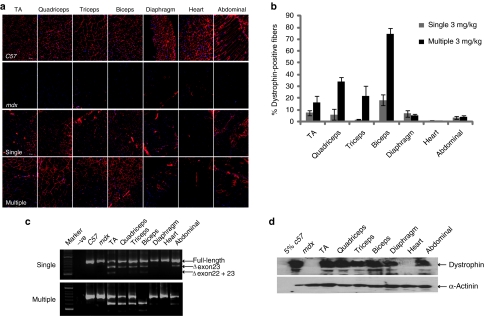
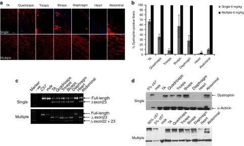
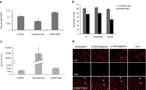
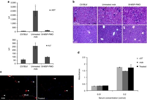
Splice modulation using antisense oligonucleotides (AOs) has been shown to yield targeted exon exclusion to restore the open reading frame and generate truncated but partially functional dystrophin protein. This has been successfully demonstrated in dystrophin-deficient mdx mice and in Duchenne muscular dystrophy (DMD) patients. However, DMD is a systemic disease; successful therapeutic exploitation of this approach will therefore depend on effective systemic delivery of AOs to all affected tissues. We have previously shown the potential of a muscle-specific/arginine-rich chimeric peptide-phosphorodiamidate morpholino (PMO) conjugate, but its long-term activity, optimized dosing regimen, capacity for functional correction and safety profile remain to be established. Here, we report the results of this chimeric peptide-PMO conjugate in the mdx mouse using low doses (3 and 6 mg/kg) administered via a 6 biweekly systemic intravenous injection protocol. We show 100% dystrophin-positive fibers and near complete correction of the dystrophin transcript defect in all peripheral muscle groups, with restoration of 50% dystrophin protein over 12 weeks, leading to correction of the DMD pathological phenotype and restoration of muscle function in the absence of detectable toxicity or immune response. Chimeric muscle-specific/cell-penetrating peptides therefore represent highly promising agents for systemic delivery of splice-correcting PMO oligomers for DMD therapy.


Figures

Figure 1
Figure 1
Systemic administration of B-MSP-PMO conjugate at single and multiple 3 mg/kg doses in mdx mice. Dystrophin expression following single and six biweekly multiple 3 mg/kg injections of the B-MSP-PMO conjugate in adult mdx mice. (a) Immunostaining of muscle tissue cross-sections to detect dystrophin protein expression and localization in mdx mice treated with single injection (the third panel, single) and multiple injections of B-MSP-PMO conjugates (bottom panel, multiple). Data from control normal C57BL6 and untreated mdx mice were shown (first and the second panel, respectively). Muscle tissues analyzed were from tibialis anterior (TA), quadriceps, triceps, biceps, abdominal wall (abdominal), and diaphragm and heart muscles. Bar = 200 µm. (b) Percent of dystrophin-positive fibers in analyzed muscle tissue cross-sections from mdx mice treated with single and multiple 3 mg/kg B-MSP-PMO conjugate doses (six sections per tissue/muscle analyzed). (c) RT-PCR analysis to detect dystrophin exon-skipping transcripts in the treated tissues with B-MSP-PMO. Unskipped and skipped transcript products as indicated (exon-skipped bands indicated by Δexon23—for exon 23 deleted; Δexon22 + 23—for exons 22 and 23 deleted). (d) Western blot for detecting the level of dystrophin protein restored in the analyzed tissues from mdx mice treated with multiple 3 mg/kg B-MSP-PMO conjugate doses compared with C57BL6 and untreated mdx control mice. 50 µg protein was loaded for each sample except for C57BL6 control lane where 2.5 µg of protein was loaded. α-Actinin was used as a loading control (six mice in each group). MSP, muscle-specific heptapeptide; PMO, phosphorodiamidate morpholino; RT, reverse transcriptase.
Figure 2
Figure 2
Long-term study of B-MSP-PMO conjugate at the dose of 6 mg/kg following six biweekly injections in mdx mice. Dystrophin exon skipping and protein expression following single and multiple systemic doses of the B-MSP-PMO conjugate in adult mdx mice. (a) Immunohistochemistry to detect dystrophin expression in muscle cross-sections from mdx mice treated with B-MSP-PMO at single 6 mg/kg dose (upper panel, single) and multiple 6 mg/kg injections (lower panel, multiple). Data from control normal C57BL6 and untreated mdx mice not shown. Muscle tissues analyzed were from tibialis anterior (TA), quadriceps, triceps, biceps, abdominal wall (abdominal), diaphragm and heart muscles. Bar = 200 µm. (b) Percent of dystrophin-positive fibers in the analyzed tissues from treated mdx mice. Results show 100% dystrophin-positive fibers observed in all the indicated muscles except heart (six sections per tissue/muscle analyzed). (c) RT-PCR to detect the dystrophin exon-skipping products in treated mdx mouse muscle groups as shown (exon-skipped bands indicated by Δexon23—for exon 23 deleted; Δexon22 + 23—for exons 22 and 23 deleted). (d) Western blot for detection of dystrophin protein in the indicated muscle groups from treated mdx mice compared with C57BL6 and untreated mdx control mice. Top panel is for single injection and the bottom one represents multiple injections. Twenty-five microgram protein was loaded for each sample except for C57BL6 control lane where a corresponding amount of protein was loaded for different treatments as indicated. α-Actinin was used as the loading control (six mice in each group). MSP, muscle-specific heptapeptide; PMO, phosphorodiamidate morpholino; RT, reverse transcriptase.
Figure 3
Figure 3
Functional and phenotypic correction in mdx mice following treatment with the B-MSP-PMO conjugate at six biweekly 6 mg/kg doses. (a) Muscle function was assessed using a functional grip strength test to determine the physical improvement of B-MSP-PMO-treated mdx mice. Significant force recovery was detected in treated mdx mice compared with untreated controls(t-test, *P < 0.05; six mice each group). (b) Evaluation of the numbers of centrally nucleated myofibers in TA, quadriceps, and biceps muscles following repeated B-MSP-PMO treatment compared with the corresponding untreated mdx muscles. Data show a significant decrease in the number of centrally nucleated myofibers in treated mdx muscles compared with untreated controls (t-test, P < 0.05; six sections per tissue/muscle analyzed; six mice each group). (c) Measurement of serum creatine kinase (CK) levels as an index of ongoing muscle membrane instability in treated mdx mice compared with mdx control mice. Data show a significant fall in the serum CK levels in mdx mice treated with B-MSP-PMO compared with untreated age-matched mdx controls (t-test, P < 0.05; six mice each group). (d) Restoration of the dystrophin-associated protein complex (DAPC) in mdx mice with repeated B-MSP-PMO treatment at 6 mg/kg was studied to assess dystrophin function and recovery of normal myoarchitecture. DAPC protein components β-dystroglycan, α-sarcoglycan, and nNOS were detected by immunostaining in serial tissue cross-sections of TA muscles from treated mdx mice compared with untreated mdx mice and normal C57BL6 mice (arrow indicated identical muscle fibers). Bar = 200 µm. MSP, muscle-specific heptapeptide; nNOS, neuronal nitric oxide synthase; PMO, phosphorodiamidate morpholino.
Figure 4
Figure 4
Investigation of systemic immunotoxicity in mdx mice treated with the multiple B-MSP-PMO conjugate administration. (a) Measurement of serum levels of aspartate aminotransferase (AST) and alanine aminotransferase (ALT) enzymes in treated mdx mice compared with untreated mdx mice. Data show a improved pathological parameters in B-MSP-PMO-treated mdx mice compared with untreated controls with significantly lower serum levels of both enzymes (t-test, *P < 0.05; six mice each group). (b) Hematoxylin and eosin staining of kidney (upper panel) and liver (lower panel) tissues sections from treated mdx mice with B-MSP-PMO, untreated mdx mice, and C57BL6 normal controls. Bar = 200 µm. Tissue sections from renal cortex show normal glomerular and tubule tissue architecture in B-MSP-PMO treated and control mice. No difference was observed for B-MSP-PMO treated mdx mice and untreated mdx controls in liver sections. (c) Detection of CD3+ T lymphocytes in the diaphragms of treated and untreated mdx mice. Bar = 100 µm. Arrows indicate the T lymphocytes detected by CD3 mouse monoclonal antibody. (d) ELISA results to detect specific antibody against the B-MSP-PMO compound in the serum from the treated mdx mice at 6 mg/kg biweekly dosing regimens. No difference was observed between treated and untreated mdx mice (six mice each group). ELISA, enzyme-linked immunosorbent assay; MSP, muscle-specific heptapeptide; PMO, phosphorodiamidate morpholino.

Similar articles

Cited by

References

    1. Wood MJ, Gait MJ., and, Yin H. RNA-targeted splice-correction therapy for neuromuscular disease. Brain. 2010;133 Pt 4:957–972. - PubMed
    1. Aartsma-Rus A, Janson AA, Kaman WE, Bremmer-Bout M, den Dunnen JT, Baas F, et al. Therapeutic antisense-induced exon skipping in cultured muscle cells from six different DMD patients. Hum Mol Genet. 2003;12:907–914. - PubMed
    1. Aartsma-Rus A, Janson AA, Kaman WE, Bremmer-Bout M, van Ommen GJ, den Dunnen JT, et al. Antisense-induced multiexon skipping for Duchenne muscular dystrophy makes more sense. Am J Hum Genet. 2004;74:83–92. - PMC - PubMed
    1. Aartsma-Rus A, Kaman WE, Weij R, den Dunnen JT, van Ommen GJ., and, van Deutekom JC. Exploring the frontiers of therapeutic exon skipping for Duchenne muscular dystrophy by double targeting within one or multiple exons. Mol Ther. 2006;14:401–407. - PubMed
    1. Alter J, Lou F, Rabinowitz A, Yin H, Rosenfeld J, Wilton SD, et al. Systemic delivery of morpholino oligonucleotide restores dystrophin expression bodywide and improves dystrophic pathology. Nat Med. 2006;12:175–177. - PubMed

Publication types

LinkOut - more resources