Inraoperative and Histological Visualization of Disrupted Vulnerable Plaques following Diagnostic Angiography of Moderate Carotid Stenosis
- PMID: 20700419
- PMCID: PMC2911585
- DOI: 10.4061/2010/602642
Inraoperative and Histological Visualization of Disrupted Vulnerable Plaques following Diagnostic Angiography of Moderate Carotid Stenosis
Abstract
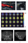
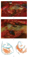
Background. Digital subtraction angiography (DSA) remains an important tool for diagnosis of carotid stenosis but is associated with risk for periprocedural complications. This is the first report of direct intraoperative and histolopathologic visualization of DSA-related carotid plaque disruption. Case. A 64-year-old man diagnosed to have a 60% right carotid stenosis received diagnostic DSA for therapeutic decision-making. He developed transient left hand numbness and weakness immediately after the procedure. Intraoperative imaging during carotid endarterectomy revealed a fragile plaque with sharp surface laceration and intraplaque hemorrhage at the bifurcation. Microscopy of the specimen demonstrated a large atheromatous plaque with fibrous hypertrophy and intraplaque hemorrhage filled with recent hemorrhagic debris. Conclusion. The visualized carotid lesion was more serious than expected, warning the danger of embolization or occlusion associated with the catheter maneuvers. Thus the highest level of practitioner training and technical expertise that ensures precise assessment of plaque characteristics should be encouraged.
Figures


Similar articles
-
Intraplaque Hemorrhage and the Plaque Surface in Carotid Atherosclerosis: The Plaque At RISK Study (PARISK).AJNR Am J Neuroradiol. 2015 Nov;36(11):2127-33. doi: 10.3174/ajnr.A4414. Epub 2015 Aug 6. AJNR Am J Neuroradiol. 2015. PMID: 26251429 Free PMC article. Clinical Trial.
-
The detection of carotid plaque rupture caused by intraplaque hemorrhage by serial high-resolution magnetic resonance imaging: a case report.Surg Neurol. 2008 Dec;70(6):634-9; discussion 639. doi: 10.1016/j.surneu.2007.06.070. Epub 2008 Jan 22. Surg Neurol. 2008. PMID: 18207497
-
Detection of carotid artery stenosis using histological specimens: a comparison of CT angiography, magnetic resonance angiography, digital subtraction angiography and Doppler ultrasonography.Acta Neurochir (Wien). 2016 Aug;158(8):1505-14. doi: 10.1007/s00701-016-2842-0. Epub 2016 Jun 2. Acta Neurochir (Wien). 2016. PMID: 27255656
-
Contemporary carotid imaging: from degree of stenosis to plaque vulnerability.J Neurosurg. 2016 Jan;124(1):27-42. doi: 10.3171/2015.1.JNS142452. Epub 2015 Jul 31. J Neurosurg. 2016. PMID: 26230478 Review.
-
[Diagnostic imaging of carotid stenosis: ultrasound, magnetic resonance imaging, and computed tomography angiography].Nihon Geka Gakkai Zasshi. 2011 Nov;112(6):371-6. Nihon Geka Gakkai Zasshi. 2011. PMID: 22165710 Review. Japanese.
Cited by
-
Intraoperative Laser Speckle Contrast Imaging For Real-Time Visualization of Cerebral Blood Flow in Cerebrovascular Surgery: Results From Pre-Clinical Studies.Sci Rep. 2020 May 6;10(1):7614. doi: 10.1038/s41598-020-64492-5. Sci Rep. 2020. PMID: 32376983 Free PMC article.
-
Dysregulation of miR-637 serves as a diagnostic biomarker in patients with carotid artery stenosis and predicts the occurrence of the cerebral ischemic event.Bioengineered. 2021 Dec;12(1):8658-8665. doi: 10.1080/21655979.2021.1988369. Bioengineered. 2021. PMID: 34606407 Free PMC article.
References
-
- Toole JF. Endarterectomy for asymptomatic carotid artery stenosis: Executive Committee for Asymptomatic Carotid Atherosclerosis Study. Journal of the American Medical Association. 1995;273(18):1421–1428. - PubMed
-
- Barnett HJM, Taylor DW, Eliasziw M, et al. Benefit of carotid endarterectomy in patients with symptomatic moderate or severe stenosis. North American Symptomatic Carotid Endarterectomy Trial Collaborators. The New England Journal of Medicine. 1998;339(20):1415–1425. - PubMed
-
- Warlow C, Farrell B, Fraser A, Sandercock P, Slattery J. Randomised trial of endarterectomy for recently symptomatic carotid stenosis: final results of the MRC European Carotid Surgery Trial (ECST) The Lancet. 1998;351(9113):1379–1387. - PubMed
-
- Rothwell PM, Goldstein LB. Carotid endarterectomy for asymptomatic carotid stenosis: asymptomatic carotid surgery trial. Stroke. 2004;35(10):2425–2427. - PubMed
-
- Nighoghossian N, Derex L, Douek P. The vulnerable carotid artery plaque: current imaging methods and new perspectives. Stroke. 2005;36(12):2764–2772. - PubMed
Publication types
LinkOut - more resources
Full Text Sources

