. 2010 Aug 5;6(8):e1001054.
doi: 10.1371/journal.pgen.1001054.

A developmental timing switch promotes axon outgrowth independent of known guidance receptors

Affiliations

A developmental timing switch promotes axon outgrowth independent of known guidance receptors

Katherine Olsson-Carter et al. PLoS Genet. .

Abstract

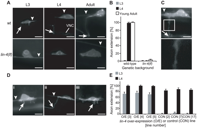
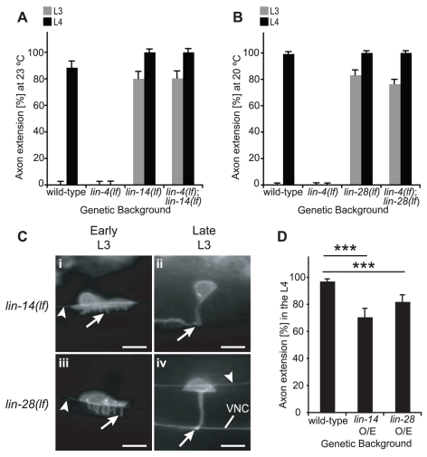
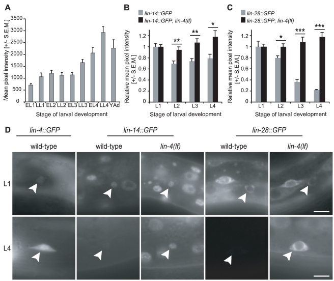
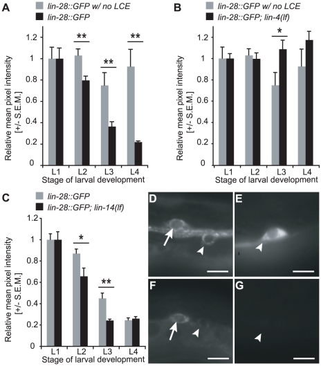
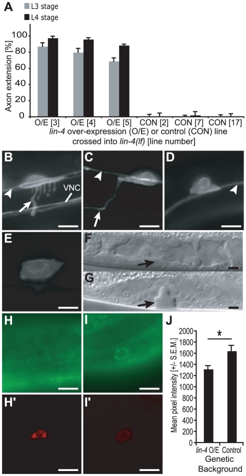
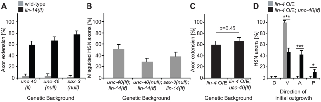
To form functional neuronal connections, axon outgrowth and guidance must be tightly regulated across space as well as time. While a number of genes and pathways have been shown to control spatial features of axon development, very little is known about the in vivo mechanisms that direct the timing of axon initiation and elongation. The Caenorhabditis elegans hermaphrodite specific motor neurons (HSNs) extend a single axon ventrally and then anteriorly during the L4 larval stage. Here we show the lin-4 microRNA promotes HSN axon initiation after cell cycle withdrawal. Axons fail to form in lin-4 mutants, while they grow prematurely in lin-4-overexpressing animals. lin-4 is required to down-regulate two inhibitors of HSN differentiation--the transcriptional regulator LIN-14 and the "stemness" factor LIN-28--and it likely does so through a cell-autonomous mechanism. This developmental switch depends neither on the UNC-40/DCC and SAX-3/Robo receptors nor on the direction of axon growth, demonstrating that it acts independently of ventral guidance signals to control the timing of HSN axon elongation.


Conflict of interest statement

The authors have declared that no competing interests exist.

Figures

Figure 1
Figure 1. lin-4(lf) displays delayed HSN axon extension.
(A) In late L3 wild-type (wt) animals (top), HSNs extended multiple neurites (arrow) in the ventral direction (left). By early L4 (middle) and in the adult (right), wild-type animals extended a single HSN axon ventrally and then anteriorly. VNC: ventral nerve cord. In L3, L4, and adult lin-4(lf) animals (bottom), no neurites or axons were typically seen. (B) Percentage of L3, L4, or adult wild-type or lin-4(lf) animals in which HSN axons had completed their anterior turn. n≥100 for each time point. (C) Image of one of the few lin-4(lf) adult animals with axon outgrowth (top). When the region contained by the box is magnified (bottom), variation in axon diameter is clearly visible. (D) Cell-autonomous over-expression of the lin-4 O/E construct led to precocious neurite outgrowth in early L3 (i) and axon elongation by late L3 (ii), while over-expression of the control construct resulted in neurite outgrowth, but not axon elongation, by late L3 (iii). (E) Percentage of L3- or L4-stage animals with axon extension in lin-4 over-expression (O/E) or control (CON) lines. All lin-4 O/E (Lines 3, 4, and 5) or control (Lines 2, 7, and 17) strains contained the integrated unc-86::myr-GFP reporter to visualize axon outgrowth. n≥50 for each time point. In A, C and D, the arrowheads point to the PLM axon, scale bars represent 5 µm, and anterior is depicted to the left and ventral is down. In A (wt L3), Di and Diii, the arrow points to one of several neurites, and in A (wt L4 and Adult), C, and Dii, it refers to the HSN axon's anterior turn. For B and E, error bars represent standard error of proportion.
Figure 2
Figure 2. lin-14 and lin-28 control the timing of HSN axon extension.
(A) Single mutant lin-14(lf) animals extended axons precociously, and in double lin-4(lf); lin-14(lf) mutants, lin-14(lf) was sufficient to suppress the lin-4(lf) retarded phenotype at the restrictive temperature (23°C). n≥50 for all time points. (B) lin-28(lf) animals displayed precocious axon outgrowth, and lin-28(lf) completely suppressed the lin-4(lf) retarded phenotype. n≥100 for all conditions. (C) Representative HSNs from lin-14(lf) (i–ii) or lin-28(lf) (iii–iv) early L3 animals with precocious outgrowth of multiple ventral neurites (arrow, i,iii) or late L3-stage animals with precocious axon outgrowth (arrow, ii,iv). Arrowhead: the PLM axon. VNC: ventral nerve cord. Animals are depicted with anterior to the left and ventral down, and scale bars represent 5 µm. (D) Over-expression of lin-14 or lin-28 led to a significant delay in axon extension during the L4 stage. ***: p<0.001 for the difference between wild-type and either lin-14 or lin-28 O/E using the two-sample z-test. n≥50 for each strain. For (A, B, D), error bars represent standard error of proportion.
Figure 3
Figure 3. lin-4 and its targets lin-14 and lin-28 are expressed reciprocally during larval development.
(A) In an integrated lin-4::GFP reporter strain, GFP is up-regulated in the HSNs during larval development. The y-axis depicts raw average pixel intensity values, and n = 10 for each time point. Note that the change in GFP expression levels between L1 and Adult stages exceeded the dynamic range of the camera, and at the set exposure time, only pixel intensity values for L1, L2, and L3 were always within the linear range. Some images acquired for late L4 and young adult stages were oversaturated, and thus the changes in pixel intensity after L3 are likely to be under-representations of the true fold increases in GFP expression. EL1–4: Early Larval Stage 1–4. LL1–4: Late Larval Stage 1–4. YAd: Young Adult. (B) GFP expression from an integrated lin-14::GFP reporter was down-regulated in the HSNs by the L2 stage, while in lin-4(lf) mutants, GFP expression was maintained. **p-values≤0.01 (0.005 for L2 and 0.003 for L3). *p-value = 0.015. (C) A lin-28::GFP reporter strain (Line 10-2) was down-regulated in the HSNs during L2 and L3 stages. When it was crossed into lin-4(lf), GFP expression persisted throughout larval development. *p-value = 0.012. ***p-values<0.001 (2.5×10⁻⁵ for L3 and 6.3×10⁻⁷ for L4). For B and C, n≥10 for each time point. Note that for each strain, pixel intensity values for L2, L3, and L4 were normalized to average L1 values and do not represent absolute fluorescence intensities. In addition, the lin-14::GFP strain expressed GFP more weakly than the lin-28 reporter during the L1, and thus a much smaller decrease in relative pixel intensity was required for complete down-regulation of the lin-14::GFP transgene. p-values for differences in relative pixel intensity were obtained using the two-sample t-test. In (A–C), all error bars represent standard error of the mean (S.E.M.). (D) Representative images of HSNs from L1 (top) and L4 (bottom) animals bearing lin-4, lin-14, or lin-28 GFP reporters in wild-type or lin-4(lf) backgrounds as noted. Arrowhead: HSN. Scale bars represent 5 µm, and anterior is to the left and ventral is down.
Figure 4
Figure 4. Regulation of lin-28 by lin-4 and lin-14 changes over developmental time.
(A) Relative GFP levels were higher for lin-28::GFPΔLCE (Line 5-1) lacking the LCE than for lin-28::GFP (Line 10-2) with an intact 3′UTR at the L2, L3, and L4 stages. **p-values≤0.01 (0.005 for L2, 0.01 for L3, and 0.002 for L4). (B) Relative GFP intensity was lower at the L3 stage in lin-28::GFPΔLCE (Line 5-1) lacking the LCE than in lin-28::GFP (Line 10-2) crossed into lin-4(lf). *: p-value = 0.030. For A–B, changes in LIN-28::GFP levels across larval development in animals lacking the LCE or lin-4 exceeded the dynamic range of the camera. At the set exposure time, some images acquired for late L4 were oversaturated. n≥10 for each strain and time point. (C) Relative lin-28::GFP (Line 10-2) expression was lower in L2- and L3-stage HSNs in lin-14(lf) compared to wild-type at 23°C. n≥9 for each lin-28::GFP condition and n≥18 for each lin-14(lf); lin-28::GFP condition. *: p-value = 0.019. **: p-value = 0.001. For A–C, data were normalized to L1 values for each strain, p-values for differences in relative pixel intensity were obtained using the two-sample t-test, and error bars represent S.E.M. (D,E) Representative L1- (D) and L4-stage (E) HSNs from lin-28::GFPΔLCE (Line 5-1). (F,G) Representative L1- (F) and L4-stage (G) HSNs from lin-28::GFP (Line 10-2); lin-14(lf). For (D–G), scale bars represent 5 µm, and anterior is left and ventral is down. Arrowhead: HSN. Arrow: CAN neuron.
Figure 5
Figure 5. lin-4 regulates HSN axon extension cell autonomously.
(A) Strains containing the lin-4(lf) mutation and the integrated unc-86::myr-GFP reporter were crossed into the lin-4 over-expression (Lines 3, 4, and 5) or control (Lines 2, 7, and 17) strains, and L3 and L4 animals were scored for axon outgrowth. The percentage of animals with axon extension in at least one HSN is shown, with n≥50 for each stage. Error bars represent standard error of proportion. (B,C) In animals with the lin-4 over-expression construct, multiple ventral neurites were detected in HSNs in early L3 (B, arrow), and axon outgrowth was visible in the mid-to-late L4 (C, arrow). VNC: ventral nerve cord. (D,E) In animals expressing the control construct, no neurites or axons were detectable at the L3 (D) or L4 (E) stages. In (B–D), arrowhead: PLM axon. (F) In the animal depicted in C, the lin-4(lf) vulvaless phenotype was not rescued and vulva development was not observed (arrow). (G) Developing vulva (arrow) from wild-type mid-to-late L4 animal. (H–J) The unc-86 promoter was used to drive over-expression of the lin-4 hairpin or a control lacking the mature lin-4 sequence in the HSNs. Representative lin-4 over-expression (O/E) (Line 4) and control (Line 7) strains were crossed into a lin-28::GFP sensor carrying its native 3'UTR (Line 10-2). In both the lin-4 O/E and control lines, HSNs inheriting the transgenes expressed the dsRED2 marker. (H–I′) Representative images of HSNs expressing either the lin-4 O/E (H and H′) or control (I and I′) construct, acquired with GFP(BP) (H and I) or TRITC (H′ and I′) filters. The scale bars in B–I′ represent 5 µm, and anterior is to the left and ventral is down. (J) The mean pixel intensity in 10 L2-stage HSNs from the lin-4 O/E and control strains. *: p-value = 0.021 using a two-sample t-test for the difference between two means. Error bars represent S.E.M.
Figure 6
Figure 6. Precocious axon outgrowth is not dependent on proper guidance.
(A) Strains containing strong loss-of-function (lf) or null alleles of unc-40 and sax-3 were scored for timing of axon outgrowth at the L3 larval stage in the presence or absence of lin-14(lf). At the restrictive temperature (23°C), lin-14(lf) animals continued to extend axons precociously in unc-40(lf), unc-40(null), or sax-3(null) mutant backgrounds. By contrast, single unc-40 or sax-3 mutants did not display marked outgrowth at the L3 stage. (B) HSN axons scored in A were categorized as misguided if they failed to extend initially in the ventral direction. (C) L3-stage animals expressing an integrated lin-4 over-expression (O/E) construct displayed precocious axon extension. There was no statistically significant change in outgrowth (p = 0.45) in the presence of unc-40(lf). (D) The predominant initial direction of growth for HSN axons scored in (C) were categorized as dorsal (D), ventral (V), anterior (A), or posterior (P). *p-value = 0.038. ***p-values are both <0.0001. For (A,C), the percentage of animals with axon extension in at least one HSN is shown, with n≥50 for each stage. For (C,D), p-values were determined using the two-sample z-test. For (A–D), error bars represent standard error of proportion.

Similar articles

Cited by

References

    1. Polleux F, Ince-Dunn G, Ghosh A. Transcriptional regulation of vertebrate axon guidance and synapse formation. Nat Rev Neurosci. 2007;8:331–340. - PubMed
    1. O'Donnell M, Chance RK, Bashaw GJ. Axon growth and guidance: receptor regulation and signal transduction. Annu Rev Neurosci. 2009;32:383–412. - PMC - PubMed
    1. Kennedy TE. Cellular mechanisms of netrin function: long-range and short-range actions. Biochem Cell Biol. 2000;78:569–575. - PubMed
    1. Butler SJ, Tear G. Getting axons onto the right path: the role of transcription factors in axon guidance. Development. 2007;134:439–448. - PubMed
    1. Adler CE, Fetter RD, Bargmann CI. UNC-6/Netrin induces neuronal asymmetry and defines the site of axon formation. Nat Neurosci. 2006;9:511–518. - PMC - PubMed

Publication types

MeSH terms