. 2010 Aug 5;5(8):e11973.
doi: 10.1371/journal.pone.0011973.

Evidence for the existence of secretory granule (dense-core vesicle)-based inositol 1,4,5-trisphosphate-dependent Ca2+ signaling system in astrocytes

Affiliations

Evidence for the existence of secretory granule (dense-core vesicle)-based inositol 1,4,5-trisphosphate-dependent Ca2+ signaling system in astrocytes

Yong Suk Hur et al. PLoS One. .

Abstract

Background: The gliotransmitters released from astrocytes are deemed to play key roles in the glial cell-neuron communication for normal function of the brain. The gliotransmitters, such as glutamate, ATP, D-serine, neuropeptide Y, are stored in vesicles of astrocytes and secreted following the inositol 1,4,5-trisphosphate (IP3)-induced intracellular Ca2+ releases. Yet studies on the identity of the IP3-dependent intracellular Ca2+ stores remain virtually unexplored.

Principal findings: We have therefore studied the potential existence of the IP3-sensitive intracellular Ca2+ stores in the cytoplasm of astrocytes using human brain tissue samples in contrast to cultured astrocytes that had primarily been used in the past. It was thus found that secretory granule marker proteins chromogranins and secretogranin II localize in the large dense core vesicles of astrocytes, thereby confirming the large dense core vesicles as bona fide secretory granules. Moreover, consistent with the major IP3-dependent intracellular Ca2+ store role of secretory granules in secretory cells, secretory granules of astrocytes also contained all three (types 1, 2, and 3) IP3R isoforms.

Significance: Given that the secretory granule marker proteins chromogranins and secretogranin II are high-capacity, low-affinity Ca2+ storage proteins and chromogranins interact with the IP3Rs to activate the IP3R/Ca2+ channels, i.e., increase both the mean open time and the open probability of the channels, these results imply that secretory granules of astrocytes function as the IP3-sensitive intracellular Ca2+ store.


Conflict of interest statement

Competing Interests: The authors have declared that no competing interests exist.

Figures

Figure 1
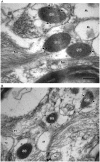
Figure 1. Electron micrographs showing the secretory granule-like vesicles (large dense-core vesicles) in astrocytes of brain tissues.
Human brain tissues were examined by electron microscope and secretory granule-like vesicles (large dense-core vesicles) of astrocytes were shown. SG, secretory granule-like vesicles; ax, axon; fm, filament. Bar = 200 nm.
Figure 2
Figure 2. Immunogold electron microscopy showing the localization of CGB and SgII in secretory granule-like vesicles (large dense-core vesicles) in astrocytes of brain tissues.
Astrocytes from human brain tissues were immunolabeled for CGB (A) and SgII (B) (15 nm gold) with the affinity purified CGB and SgII antibodies, respectively. The CGB- or SgII-labeling gold particles (indicated by arrows) were primarily localized in the secretory granule-like vesicles (SG) with some in the endoplasmic reticulum (er), but not in the mitochondria (M). In the control experiments without the primary antibodies no gold particles were seen in the secretory granule-like vesicles (not shown). Bar = 200 nm.
Figure 3
Figure 3. Localization of glial fibrillary acidic protein, CGB, and SgII in astrocytes of brain tissues.
Expression of glial fibrillary acidic proteins in the intermediate filaments of astrocytes that contain secretory granules was examined by double immunogold labeling using the antibodies specific for GFAP and either CGB (A) or SgII (B). (A) The GFAP-labeling gold particles (10 nm) and the CGB-labeling gold particles (15 nm) are marked by black and white arrows, respectively. Notice that the GFAP-labeling gold particles are exclusively localized in the filaments (fm) whereas the CGB-labeling particles are limited to secretory granules (SG). (B) The GFAP-labeling gold particles (10 nm) and the SgII-labeling gold particles (15 nm) are marked by black and white arrows, respectively. Again the GFAP-labeling gold particles are exclusively localized in the filaments (fm) whereas the SgII-labeling gold particles are localized in secretory granules (SG), but not in mitochondria (M) and axon (ax). Bar = 200 nm.
Figure 4
Figure 4. Immunogold electron microscopy showing the localization of IP3R1 in secretory granules in astrocytes.
Astrocytes from human brain tissues were immunolabeled for IP3R1 (15 nm gold) with the affinity purified IP3R1 antibody (A and B). The IP3R1-labeling gold particles (indicated by arrows) were primarily localized in the membranes of secretory granules (SG) with some in the endoplasmic reticulum (see A), but not in the mitochondria (M). In the control experiments without the primary antibody no gold particles were seen in secretory granules (not shown). fm, filament. Bar = 200 nm.
Figure 5
Figure 5. Immunogold electron microscopy showing the localization of IP3R2 in secretory granules in astrocytes.
Astrocytes from human brain tissues were immunolabeled for IP3R2 (15 nm gold) with the affinity purified IP3R2 antibody (A and B). The IP3R2-labeling gold particles (indicated by arrows) were primarily localized in the membranes of secretory granules (SG) with some in the endoplasmic reticulum (see A), but not in the mitochondria (M). In the control experiments without the primary antibody no gold particles were seen in secretory granules (not shown). ax, axon; fm, filament. Bar = 200 nm.
Figure 6
Figure 6. Immunogold electron microscopy showing the localization of IP3R3 in secretory granules in astrocytes.
Astrocytes from human brain tissues were immunolabeled for IP3R3 (15 nm gold) with the affinity purified IP3R3 antibody (A and B). The IP3R3-labeling gold particles (indicated by arrows) were primarily localized in the membranes of secretory granules (SG) with some in the endoplasmic reticulum (see B), but not in the mitochondria (M). In the control experiments without the primary antibody no gold particles were seen in secretory granules (not shown). ax, axon; fm, filament. Bar = 200 nm.
Figure 7
Figure 7. A model showing the IP3-induced Ca2+ mobilization from secretory granules and the secretory processes of astrocytes.
The tetrameric IP3R/Ca2+ channels are shown in red and blue columns while chromogranins A and B are shown in open and hatched circles, respectively. Only can chromogranin B, which interacts with CGA to form a CGA-CGB heterodimer at the pH of ER, couple to the tetrameric IP3Rs in the ER whereas both chromogranins A and B, which form a CGA2CGB2 heterotetrameric complex at the acidic intragranular pH , couple to the tetrameric IP3Rs in secretory granules , , . Stimuli at the cell surface (1) will lead to the production of IP3 at the plasma membrane, which will serve as the first signal to induce the IP3-dependent Ca2+ release from intracellular Ca2+ stores in the cytoplasm. Yet each intracellular Ca2+ store will respond differently to IP3 depending on the amount of IP3 produced and the sensitivity of the IP3R/Ca2+ channels to IP3. In light of the significantly higher sensitivity of the IP3R/Ca2+ channels of secretory granules than those of the ER , secretory granules will release Ca2+ (2), ahead of the ER (3), in response to low IP3 concentrations. This Ca2+ could play essential roles in initiating secretion by synaptic-like vesicles (4) and secretory granules (5), leading to secretion of the gliotransmitters (6).

References

    1. Angulo MC, Kozlov AS, Charpak S, Audinat E. Glutamate released from glial cells synchronizes neuronal activity in the hippocampus. J Neurosci. 2004;24:6920–6927. - PMC - PubMed
    1. Perea G, Araque A. Glial calcium signaling and neuron-glia communication. Cell Calcium. 2005;38:375–382. - PubMed
    1. Volterra A, Meldolesi J. Astrocytes, from brain glue to communication elements: the revolution continues. Nat Rev Neurosci. 2005;6:626–640. - PubMed
    1. Panatier A, Theodosis DT, Mothet JP, Touquet B, Pollegioni L, et al. Glia-derived D-serine controls NMDA receptor activity and synaptic memory. Cell. 2006;125:775–784. - PubMed
    1. Montana V, Malarkey EB, Verderio C, Matteoli M, Parpura V. Vesicular transmitter release from astrocytes. Glia. 2006;54:700–715. - PubMed

Publication types

MeSH terms