. 2010 Aug 5;5(8):e11977.
doi: 10.1371/journal.pone.0011977.

The native Wolbachia endosymbionts of Drosophila melanogaster and Culex quinquefasciatus increase host resistance to West Nile virus infection

Affiliations

The native Wolbachia endosymbionts of Drosophila melanogaster and Culex quinquefasciatus increase host resistance to West Nile virus infection

Robert L Glaser et al. PLoS One. .

Abstract

Background: The bacterial endosymbiont Wolbachia pipientis has been shown to increase host resistance to viral infection in native Drosophila hosts and in the normally Wolbachia-free heterologous host Aedes aegypti when infected by Wolbachia from Drosophila melanogaster or Aedes albopictus. Wolbachia infection has not yet been demonstrated to increase viral resistance in a native Wolbachia-mosquito host system.

Methodology/principal findings: In this study, we investigated Wolbachia-induced resistance to West Nile virus (WNV; Flaviviridae) by measuring infection susceptibility in Wolbachia-infected and Wolbachia-free D. melanogaster and Culex quinquefasciatus, a natural mosquito vector of WNV. Wolbachia infection of D. melanogaster induces strong resistance to WNV infection. Wolbachia-infected flies had a 500-fold higher ID50 for WNV and produced 100,000-fold lower virus titers compared to flies lacking Wolbachia. The resistance phenotype was transmitted as a maternal, cytoplasmic factor and was fully reverted in flies cured of Wolbachia. Wolbachia infection had much less effect on the susceptibility of D. melanogaster to Chikungunya (Togaviridae) and La Crosse (Bunyaviridae) viruses. Wolbachia also induces resistance to WNV infection in Cx. quinquefasciatus. While Wolbachia had no effect on the overall rate of peroral infection by WNV, Wolbachia-infected mosquitoes produced lower virus titers and had 2 to 3-fold lower rates of virus transmission compared to mosquitoes lacking Wolbachia.

Conclusions/significance: This is the first demonstration that Wolbachia can increase resistance to arbovirus infection resulting in decreased virus transmission in a native Wolbachia-mosquito system. The results suggest that Wolbachia reduces vector competence in Cx. quinquefasciatus, and potentially in other Wolbachia-infected mosquito vectors.


Conflict of interest statement

Competing Interests: The authors have declared that no competing interests exist.

Figures

Figure 1
Figure 1. Wolbachia status of D. melanogaster and Cx. quinquefasciatus strains analyzed for susceptibility to arbovirus infection.
DNA was isolated from D. melanogaster strains Oregon R (OR) and BER1, and from Cx. quinquefasciatus (Cxq). Tetracycline-treated strains lacking Wolbachia sequences are indicated by the suffix -T. DNA sequences corresponding to the wsp gene of Wolbachia and the 12S mitochondrial gene were identified by PCR.
Figure 2
Figure 2. Wolbachia-induced resistance to WNV infection in wild-type D. melanogaster strain BER1.
The indicated pfu of WNV was injected into Wolbachia(+) BER1 and Wolbachia(−) BER1-T strains of D. melanogaster. Seven days after inoculation, the titer of WNV in each fly was measured by plaque assay. (A) The fraction of flies that became infected for each genotype at each concentration of virus, and the ID50 value for each genotype as calculated from those data, are shown. (B) The titers of WNV in infected Wolbachia(+) BER1 (X) and Wolbachia(−) BER1-T flies (O) are shown. The grey diagonal line indicates the amount of WNV inoculated per fly. The limit of detection of the plaque assay was 5 pfu/animal.
Figure 3
Figure 3. Tetracycline treatment of Wolbachia(−) Oregon R flies had little effect on susceptibility to WNV infection.
The indicated pfu of WNV was injected into untreated (OR) and tetracycline-treated (OR-T) Oregon R flies. Seven days after inoculation, the titer of WNV in each fly was measured by plaque assay. (A) The fraction of flies that became infected for each genotype at each concentration of virus, and the ID50 value for each genotype as calculated from those data, are shown. (B) The titers of WNV in untreated (X) and tetracycline-treated flies (O) are shown. The grey diagonal line indicates the amount of WNV inoculated per fly. The limit of detection of the plaque assay was 25 pfu/animal.
Figure 4
Figure 4. Wolbachia-induced resistance to CHIKV infection in D. melanogaster.
The indicated pfu of CHIKV was injected into Wolbachia(+) BER1 and Wolbachia(−) BER1-T flies. Seven days after inoculation, the titer of CHIKV in each fly was measured by plaque assay. (A) The fraction of flies that became infected for each genotype at each concentration of virus, and the ID50 value for each genotype as calculated from those data, are shown. (B) The titers of CHIKV in infected Wolbachia(+) BER1 (O) and Wolbachia(−) BER1-T flies (X) are shown. The grey diagonal line indicates the amount of CHIKV inoculated per fly. The limit of detection of the plaque assay was 25 pfu/animal.
Figure 5
Figure 5. Wolbachia did not increase resistance to LACV infection in D. melanogaster.
The indicated pfu of LACV was injected into Wolbachia(+) BER1 and Wolbachia(−) BER1-T flies, and seven days after inoculation, the titer of LACV was measured in each fly by plaque assay. (A) The fraction of flies that became infected for each genotype at each concentration of virus, and the ID50 value for each genotype as calculated from those data, are shown. (B) The titers of LACV in infected Wolbachia(+) BER1 (O) and Wolbachia(−) BER1-T flies (X) are shown. The grey diagonal line indicates the amount of CHIKV inoculated per fly. The limit of detection of the plaque assay was 10 pfu/animal.
Figure 6
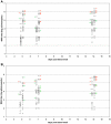
Figure 6. Titers of WNV in infected Wolbachia(+) and Wolbachia(−) Cx. quinquefasciatus mosquitoes.
Wolbachia(+) and Wolbachia(−) Cx. quinquefasciatus mosquitoes were fed a blood meal containing WNV either five generations (A) or fourteen generations (B) after tetracycline treatment. Titers of WNV in infected Wolbachia(+) mosquitoes (O) and in infected Wolbachia(−) mosquitoes (X) were measured by plaque assay 5, 7, and 14 days post blood meal. The average virus titer is indicated by a horizontal line. Mosquitoes in which virus had disseminated only to the legs (green) or that had disseminated to the legs and was transmitted in the saliva (red) are indicated by colored plus signs located next to the virus titer for that same mosquito.

Similar articles

Cited by

References

    1. Saridaki A, Bourtzis K. Wolbachia: more than just a bug in insects genitals. Curr Op Micro. 2010;13:67–72. - PubMed
    1. Anderson CL, Karr TL. Wolbachia: Evolutionary novelty in a rickettsial bacteria. BMC Evol Biol. 2001;1:10. - PMC - PubMed
    1. O'Neill SL, Giordano R, Colbert AME, Karr TL. 16S rRNA phylogenetic analysis of the bacterial endosymbionts associated with cytoplasmic incompatibility in insects. Proc Natl Acad Sci USA. 1992;89:2699–2702. - PMC - PubMed
    1. Iturbe-Ormaetxe I, O'Neill SL. Wolbachia-host interactions: connecting phenotype to genotype. Curr Opin Microbiol. 2007;10:221–224. - PubMed
    1. Bouchon D, Rigaud T, Juchault P. Evidence for widespread Wolbachia infection in isopod curstaceans: molecular identification and host feminization. Proc R Soc Lond B. 1998;265:10–81-10-90. - PMC - PubMed

Publication types

LinkOut - more resources