. 2010 Aug 5;5(8):e11980.
doi: 10.1371/journal.pone.0011980.

Oxytocin signaling in mouse taste buds

Affiliations

Oxytocin signaling in mouse taste buds

Michael S Sinclair et al. PLoS One. .

Abstract

Background: The neuropeptide, oxytocin (OXT), acts on brain circuits to inhibit food intake. Mutant mice lacking OXT (OXT knockout) overconsume salty and sweet (i.e. sucrose, saccharin) solutions. We asked if OXT might also act on taste buds via its receptor, OXTR.

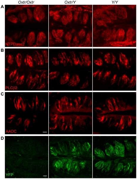
Methodology/principal findings: Using RT-PCR, we detected the expression of OXTR in taste buds throughout the oral cavity, but not in adjacent non-taste lingual epithelium. By immunostaining tissues from OXTR-YFP knock-in mice, we found that OXTR is expressed in a subset of Glial-like (Type I) taste cells, and also in cells on the periphery of taste buds. Single-cell RT-PCR confirmed this cell-type assignment. Using Ca2+ imaging, we observed that physiologically appropriate concentrations of OXT evoked [Ca2+]i mobilization in a subset of taste cells (EC50 approximately 33 nM). OXT-evoked responses were significantly inhibited by the OXTR antagonist, L-371,257. Isolated OXT-responsive taste cells were neither Receptor (Type II) nor Presynaptic (Type III) cells, consistent with our immunofluorescence observations. We also investigated the source of OXT peptide that may act on taste cells. Both RT-PCR and immunostaining suggest that the OXT peptide is not produced in taste buds or in their associated nerves. Finally, we also examined the morphology of taste buds from mice that lack OXTR. Taste buds and their constituent cell types appeared very similar in mice with two, one or no copies of the OXTR gene.

Conclusions/significance: We conclude that OXT elicits Ca2+ signals via OXTR in murine taste buds. OXT-responsive cells are most likely a subset of Glial-like (Type I) taste cells. OXT itself is not produced locally in taste tissue and is likely delivered through the circulation. Loss of OXTR does not grossly alter the morphology of any of the cell types contained in taste buds. Instead, we speculate that OXT-responsive Glial-like (Type I) taste bud cells modulate taste signaling and afferent sensory output. Such modulation would complement central pathways of appetite regulation that employ circulating homeostatic and satiety signals.


Conflict of interest statement

Competing Interests: The authors have declared that no competing interests exist.

Figures

Figure 1
Figure 1. Taste buds selectively express Oxytocin Receptor (OXTR).
A. RT-PCR for OXTR (upper) and β-actin (lower) on taste epithelium from four areas (va, vallate; fo, foliate; ff, fungiform; pa, palate), non-taste lingual epithelium (nt), brain (br) and water (−). Arrows mark predicted bands (OXTR, 187 bp; β-actin, 328 bp). B. Real-time RT-PCR performed in parallel on cDNA from vallate and nontaste epithelia (OXTR mRNA copies normalized to 1000 copies β-actin mRNA) suggests that OXTR expression is taste-selective (* = p<0.05 vallate compared to non-taste). C. Real-time RT-PCR on four fields of taste epithelium. OXTR mRNA copy number is normalized to 100 copies of PLCβ2 mRNA. Anterior epithelium (ff) and palate have more copies of OXTR mRNA per unit of taste cell mass compared to posterior taste epithelium (va, fo). Bars depict mean and s.e.m. (n = 3).
Figure 2
Figure 2. Oxytocin Receptor, is expressed in taste buds but not in Receptor or Presynaptic cells.
Cryosections of vallate (va, A) or fungiform (ff, B) papilla from OXTR-YFP mice, were immunostained for PLCβ2 (red, right) to reveal Receptor cells and taste buds, and with anti-GFP (green) to detect YFP expression. Expression of YFP is limited to taste buds. No YFP was detected in either non-sensory epithelium or underlying connective tissue (revealed by differential interference contrast microscopy in overlay at right). Higher magnification cross-sections of fungiform (ff, C) or palatal (pa, D) taste buds, from OXTR-YFP mice, immunostained for PLCβ2 (C) or Chromogranin A (D) (red) and YFP (green). OXTR is expressed in cells that are distinct from PLCβ2-positive (Receptor) cells and Chromogranin A-positive (Presynaptic) cells. In many instances, strong GFP fluorescence was detected in cells on the periphery of the taste bud (arrowheads in A–D). Scale bars are 50 µm (A,B) or 10 µm (C,D).
Figure 3
Figure 3. Oxytocin Receptor is present in Glial-like cells in taste buds.
A, B. Cryosections of vallate papilla from OXTR-YFP mice, cut horizontally (A) or vertically (B), were immunostained for NTPDase2 (red), a marker for taste glial-like cells, and for YFP (green). YFP is co-expressed in some NTPDase2-positive' cells. Membrane-localized NTPDase2 is detected in YFP-expressing cells (arrows in overlay images, right). YFP is also in cells, particularly at the periphery of the taste bud, that are not associated with NTPDase2 and thus cannot be termed Glial-like (arrowhead). Scale bars are 10 µm. C. Pools, each containing 20 taste cells of an identified type, were assayed for OXTR by RT-PCR. Receptor and Presynaptic cells were identified and harvested as GFP-positive cells from taste buds of PLCβ2-GFP (pool #2) or GAD1-GFP (pool #3) mice. To isolate glial-like cells, GFP-negative cells from PLCβ2-GFP x GAD1-GFP double transgenic mice were collected for pool #1. All pools were subjected to RT-PCR for all three diagnostic markers, NTPDase2, PLCβ2, and GAD1 to confirm their identification. OXTR was detected only in the pool (#1) of NTPDase2-expressing, GFP-negative cells. D. A typical RT-PCR experiment on 6 individual Glial-like (NTPDase2-positive) cells. OXTR is detected in 2 of the 6 cells. Arrowheads indicate expected product size in each case. Controls are water (−) or whole taste buds (tb).
Figure 4
Figure 4. OXT evokes Ca2+ responses in taste cells.
A. Pseudocolored image of fura-2 fluorescence from an isolated taste bud from a PLCβ2-GFP mouse before, during, and after Ca2+ mobilization evoked by 30 nM OXT. A cell on the periphery (arrowhead) displayed a Ca2+ response (red) that could be replicated with repeated applications of OXT. Image at right shows GFP fluorescence (green) and brightfield micrograph of the same field, overlaid. The OXT-responsive cell is GFP-negative. That is, it is not a Receptor cell. B. Ca2+ responses (F340/F380 ratio) from a taste cell similar to the one indicated in A. This cell responded robustly to 1 µM ATP but not to depolarization with 50 mM KCl (i.e. it is not a Presynaptic cell). This cell repeatedly displayed Ca2+ responses when stimulated with 30 nM OXT. C. A Presynaptic cell in a taste bud responded to ATP (1 µM) and to depolarization with 50 mM KCl, but not to 30 nM OXT.
Figure 5
Figure 5. OXT-evoked responses in taste cells are dose-dependent and can be blocked with an OXTR antagonist.
A. Trace showing OXT responses to increasing doses of OXT (10 nM to 300 nM, for 30 sec each, indicated by bars above traces). B. Concentration-response curve, based on peak responses, from 4 separate experiments. Estimated EC50 = 33 nM. N = 13 for each point except at 60 nM (N = 5) and 1 µM (N = 8). C. An example trace from an OXT-responsive cell, exposed sequentially for 30 sec each to 30 and 100 nM OXT before, during (shaded) and after incubating the preparation in 500 nM L-371,257 (L), an OXTR antagonist. Treatment with L-371,257 reversibly abolished OXT responses at both concentrations of OXT. D. Aggregate data (mean +/− s.e.m.) from 3 independent experiments show that L-371,257 significantly inhibits OXT responses in taste cells and that responses to OXT recover after washout of the inhibitor (N = 6; 2-way ANOVA with repeated measures followed by Newman-Keuls post-hoc test. ** = p<0.01 comparing Pre-L to L for each concentration of OXT; *** = p<0.001 comparing Post-L to L for each concentration of OXT). An interval of 20 min with constant flow perfusion of Tyrode buffer elapsed between successive OXT applications.
Figure 6
Figure 6. Oxytocin peptide (OXT) is not produced locally in taste tissue.
A. RT-PCR was carried out on vallate papilla (t1), two samples of vallate and foliate taste buds (t2, t3), non-taste lingual epithelium (nt, negative control), and brain (br, positive control). Primers selective for OXT (top), PLCβ2 (middle) and β-actin (bottom) were tested in parallel for each sample. The expected PCR product and size in basepairs are indicated at right. B,C. Validating the anti-OXT antibody. Coronal sections of brain from heterozygous (Oxt +/−) or knockout (Oxt −/−) mice, fixed and processed in parallel for OXT-immunostaining (red). Sections through the hypothalamus reveal a cluster of OXT-positive neurons in the Paraventricular Nucleus (PVN), in Oxt +/− but not in Oxt −/− mouse. In C, the PVN is shown at higher magnification. D. Cryosections of vallate papillae from the same Oxt +/− and Oxt −/− mice as in B,C, immunostained with the same anti-OXT antibody (green). These sections were also immunostained with anti-Tyrosine Hydroxylase (TH, red) to reveal nerve fibers. Vertical arrows point at the apical pore of two taste buds in each panel. No specific immunofluorescence for OXT was detected in taste buds, adjacent epithelium, or nerve fibers. The faint green fluorescence in these images is identical in tissues from Oxt +/− and −/− mice and thus, cannot be attributed to OXT. Certain primary and secondary antibodies, even those well-validated in other tissues, do yield similar faint non-specific background staining in taste tissue. The “gold standard” test in knockout tissue demonstrates that it is non-specific. Scale bars are 50 µm for B and 20 µm for C, D.
Figure 7
Figure 7. OXTR deficiency does not alter the morphology of taste buds.
Vallate papillae were harvested from mice of the following genotypes: wildtype (Oxtr/Oxtr), heterozygous (Oxtr/Y), or homozygous for the OXTR-YFP knock-in allele (Y/Y). The Y/Y mice have no functional allele for OXTR, and are OXTR knockouts. We immunostained sections of vallate papillae for NTPDase2 (A), PLCβ2 (B), and aromatic amino acid decarboxylase (AADC) (C). NTPDase2 and PLCβ2 are markers for Glial-like and Receptor cells, respectively. AADC is a marker for Presynaptic cells , . Sections from all three genotypes immunostained with anti-GFP (D) are shown for reference. We observed no difference in size, shape, or number of cells of each type across genotypes. Scale bars, 20 µm. Bar in C applies to A–C.

Similar articles

Cited by

References

    1. Donaldson ZR, Young LJ. Oxytocin, vasopressin, and the neurogenetics of sociality. Science. 2008;322:900–904. - PubMed
    1. Ludwig M, Leng G. Dendritic peptide release and peptide-dependent behaviours. Nat Rev Neurosci. 2006;7:126–136. - PubMed
    1. Blevins JE, Schwartz MW, Baskin DG. Evidence that paraventricular nucleus oxytocin neurons link hypothalamic leptin action to caudal brain stem nuclei controlling meal size. Am J Physiol Regul Integr Comp Physiol. 2004;287:R87–R96. - PubMed
    1. Sabatier N, Rowe I, Leng G. Central release of oxytocin and the ventromedial hypothalamus. Biochem Soc Trans. 2007;35:1247–1251. - PubMed
    1. Arletti R, Benelli A, Bertolini A. Oxytocin inhibits food and fluid intake in rats. Physiol Behav. 1990;48:825–830. - PubMed

Publication types

MeSH terms