. 2010 Aug 5;5(8):e11993.
doi: 10.1371/journal.pone.0011993.

Candida albicans AGE3, the ortholog of the S. cerevisiae ARF-GAP-encoding gene GCS1, is required for hyphal growth and drug resistance

Affiliations

Candida albicans AGE3, the ortholog of the S. cerevisiae ARF-GAP-encoding gene GCS1, is required for hyphal growth and drug resistance

Thomas Lettner et al. PLoS One. .

Abstract

Background: Hyphal growth and multidrug resistance of C. albicans are important features for virulence and antifungal therapy of this pathogenic fungus.

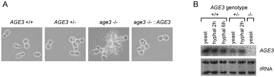
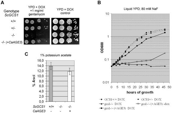
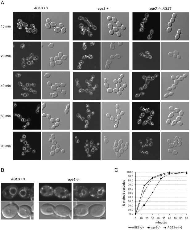
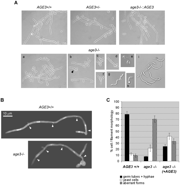
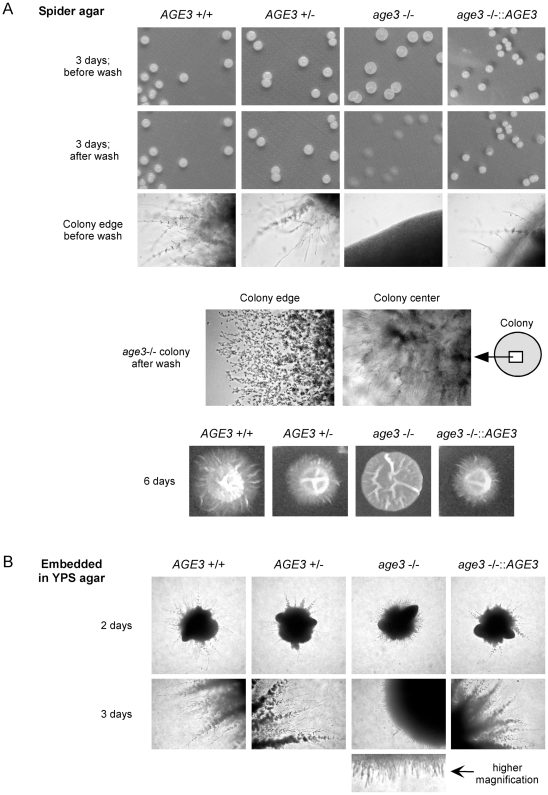
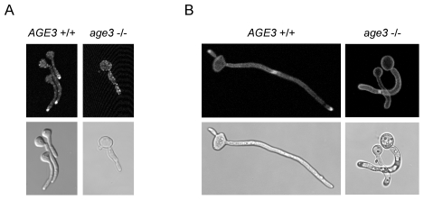
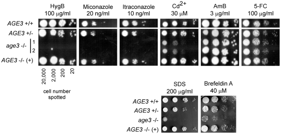
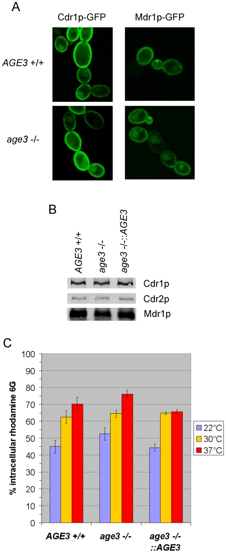
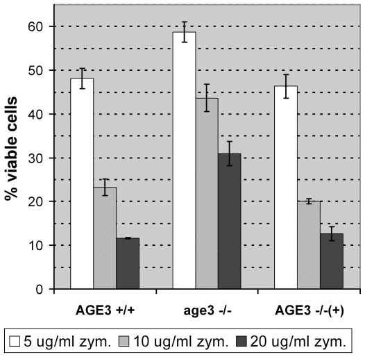
Methodology/principal findings: Here we show by phenotypic complementation analysis that the C. albicans gene AGE3 is the functional ortholog of the yeast ARF-GAP-encoding gene GCS1. The finding that the gene is required for efficient endocytosis points to an important functional role of Age3p in endosomal compartments. Most C. albicans age3Delta mutant cells which grew as cell clusters under yeast growth conditions showed defects in filamentation under different hyphal growth conditions and were almost completely disabled for invasive filamentous growth. Under hyphal growth conditions only a fraction of age3Delta cells shows a wild-type-like polarization pattern of the actin cytoskeleton and lipid rafts. Moreover, age3Delta cells were highly susceptible to several unrelated toxic compounds including antifungal azole drugs. Irrespective of the AGE3 genotype, C-terminal fusions of GFP to the drug efflux pumps Cdr1p and Mdr1p were predominantly localized in the plasma membrane. Moreover, the plasma membranes of wild-type and age3Delta mutant cells contained similar amounts of Cdr1p, Cdr2p and Mdr1p.

Conclusions/significance: The results indicate that the defect in sustaining filament elongation is probably caused by the failure of age3Delta cells to polarize the actin cytoskeleton and possibly of inefficient endocytosis. The high susceptibility of age3Delta cells to azoles is not caused by inefficient transport of efflux pumps to the cell membrane. A possible role of a vacuolar defect of age3Delta cells in drug susceptibility is proposed and discussed. In conclusion, our study shows that the ARF-GAP Age3p is required for hyphal growth which is an important virulence factor of C. albicans and essential for detoxification of azole drugs which are routinely used for antifungal therapy. Thus, it represents a promising antifungal drug target.


Conflict of interest statement

Competing Interests: The authors have declared that no competing interests exist.

Figures

Figure 1
Figure 1. Morphology of the age3Δ/Δ strain under yeast growth conditions and AGE3 gene expression.
(A) The strains SN87 (AGE3+/+), UZ22 (AGE3+/Δ), UZ45 (age3Δ/Δ) and UZ55 (age3Δ/Δ::AGE3) were grown in SD medium at 30°C to an OD600 of 0.5. Samples were inspected under the microscope (phase contrast optics). Note that the homozygous age3 mutant strain forms aggregates of yeast cells. The heterozygous and the reintegrant strain show intermediate phenotype. (B) Northern blotting of RNA isolated under yeast and hyphal growth conditions. The wild-type strain, the heterozygous and the homozygous age3 mutant strains were grown either to exponential phase in YPD or induced for hyphal growth (two and six hours) in YPD +10% bovine serum. Each 4 µg total RNA were blotted and hybridized with a 32P-labeled AGE3-specific probe. Methylene blue-stained rRNA on the blot is shown as a control for blotting efficiency.
Figure 2
Figure 2. CaAGE3 complements defects of the yeast gcs1Δ mutant.
(A) The diploid S. cerevisiae strains BY4743 (wild type), ydl226c+/− (heterozygous for GCS1), ydl226c−/− (homozygous gcs1Δ) and UZ177 (homozygous gcs1Δ transformed with the centromeric plasmid pCaAge3-2 which carries the CaAGE3 ORF under control of the doxycycline-inducible Tet promoter) were grown for 48 hours on agar plates containing 50 µg/ml doxycycline (DOX) in the presence of gentamycin (A). The number of cells spotted onto the agar was about 20,000, 2,000, 200 or 20 (from left to right). (B) Exponentially pregrown cells (strains as in A) were inoculated into 80 mM sodium fluoride in YPD +50 µg/ml DOX and incubated at 30°C for 42 hours. Samples were taken after different time points and the OD600 measured. For control, each strain was also grown without DOX. Except for strain UZ177 the growth rate was independent of the presence of DOX (not shown). However, AGE3 induction in strain UZ177 by DOX results in complementation of the growth deficiency of gcs1Δ cells. This experiment was repeated and very similar growth behaviours were observed. (C) The wild-type, the homozygous gcs1Δ strain and UZ177 were induced for sporulation on agar plates containing 1% potassium acetate and 50 µg/ml DOX. The percentage of asci formation was determined after five days incubation at 30°C. This experiment was done in triplicate. Expression of CaAGE3 complements the complete inability of the gcs1Δ/Δ strain to form asci. Note that in general the sporulation efficiency of strains derived from BY4743 is low compared to other wild-type strains.
Figure 3
Figure 3. Cells lacking AGE3 show a clear delay in endocytosis.
(A) Cells of the strains SN87 (AGE3+/+), UZ45 (age3Δ/Δ) and UZ55 (age3Δ/Δ::AGE3) were grown in YPD to exponential phase. After staining the cytoplasmic membrane with the lipophilic fluorescent dye FM4-64 at 0°C for 40 minutes, the excess dye was removed by washing. Then the cells were released for growth in YPD at 30°C to allow endocytosis to occur. Samples were taken at the indicated time points. Staining of endocytic vesicles and the vacuole was visualized by confocal fluorescence microscopy. DIC images are shown for comparison. (B) One and two cell pairs of the wild-type and the age3Δ/Δ mutant strains, respectively, which were harvested 40 minutes after release of endocytosis are shown in higher magnification. The mutant cells show stronger background staining of the cytoplasm and more bright spots compared to the wild-type cells. The vauolar membranes of the left mutant cells still have not taken up FM4-64. (C) Quantification of FM4-64 uptake into vacuolar membranes in cells of the strains mentioned above. Images of a similar experiment as described under (A) were analysed and the percentage of cells (about 100–150 in total for each strain and time point) with clearly stained vacuolar membranes determined. A similar experiment performed on another day with slightly different time points showed a very similar FM4-64 uptake kinetics.
Figure 4
Figure 4. Growth forms of the age3Δ/Δ strain after hyphal induction in YPD +10% bovine serum (A and B) or in GlcNAc medium (C).
(A) The strains SN87 (AGE3+/+), UZ22 (AGE3+/Δ), UZ45 (age3Δ/Δ) and UZ55 (age3Δ/Δ::AGE3) were induced for hyphal growth in YPD +10% bovine serum at 37°C. After five hours of growth, samples were inspected under the microscope (most pictures in phase contrast and some in Nomarsky optics). Note that the homozygous age3 mutant strain forms several distinct forms of filaments. Some of them are not true hyphal filaments. Most strikingly, a fraction of germ tubes and filaments show an untypically curved or spiral shape (images e, g, h and i). Some other filaments show constrictions between cells which are typical for pseudohyphae (i). There are also cell clusters seen which are composed of yeast-form cells (c) and others which show only poor polarized growth (d, f and b arrow head). (B) Each one true hyphal filament of the wild-type and the age3 mutant strains grown under the same conditions as in (A) were stained with Calcofluor white and DAPI to visualize septa (arrow heads) and nuclei, respectively. Compared to wild-type filaments, age3 mutant filaments are composed of shorter hyphal cells (except for the first cell following the blastospore in many cases). (C) The strains SN87 (AGE3+/+), UZ45 (age3Δ/Δ) and UZ55 (age3Δ/Δ::AGE3) were induced for hyphal growth in GlcNAc-containing medium. After three hours of growth at 37°C for each clone about 200 cells and filaments were classified as wild-type-like germ tubes and hyphae, yeast cells and aberrant forms of single cells, germ tubes or filaments (curved shape, short pseudohyphal elements, cells with thick germ tubes; as shown in (A)). The experiment was done in triplicate. The mean percentage of these morphologies and standard deviation are shown.
Figure 5
Figure 5. Defective filamentous and invasive growth of the age3Δ/Δ strain on/in solid media.
Note: All images of each row are shown in the same magnification. (A) The strains SN87 (AGE3+/+), UZ22 (AGE3+/Δ), UZ45 (age3Δ/Δ) and UZ55 (age3Δ/Δ::AGE3) were induced for filamentous growth on Spider agar at 37°C for three and six days (two sets of agar plates). The colonies were photographed and the colony edges inspected under the microscope. To try to remove the colonies after three days of growth one set of agar plates was washed with water. Only the homozygous age3 mutant colonies could be removed easily. At the edges of the mutant colonies after wash single yeast cells and very short filaments are still attached to the agar. However, weak invasive growth into the agar by age3 mutant filaments is only visible in the colony center. After six days, filamentous growth is seen macroscopically for the wild-type, the heterozygous and the reintegrant strain. The mutant strain forms flat colonies without visible filaments (as also seen after three days). (B) The strains mentioned under (A) were induced for filamentous growth by embedding in YPS agar and incubation for three days at 37°C. The homozygous age3 mutant colonies formed after two days of growth show a high rate of filamentation. However, the filaments were unable to develop further (three days) into long filaments which could invade the agar as filaments of the control strains do.
Figure 6
Figure 6. Absence of polarization of lipid rafts and actin patches in age3Δ cells.
The strains SN87 (AGE3+/+) and UZ45 (age3Δ/Δ) were induced for hyphal growth in GlcNAc medium at 37°C. After three hours samples were taken and the cell structures shown were stained and visualized under the confocal microscope. For control, pictures made with phase contrast or DIC optics are shown below the fluorescence picture. (A) At the tips of germ tubes of wild-type cells the actin patches, which were stained with Alexafluor 488-conjugated phalloidin, are highly concentrated. Most mutant cells do not show this polarization of the actin cytoskeleton. (B) Lipid rafts were stained with filipin. As expected, lipid rafts are concentrated at the hyphal tips and at septa in the wild type. Most mutant germ tubes (note that some have constrictions at the neck) and filaments show a uniform staining of the cytoplasmic membrane. Note that the figure shows only typical structures found in cultures of wild-type and mutant cells. However, a low fraction of mutant cells and filaments (see Fig. 4C) showed a wild type-like staining patterns. Also for the reintegrant strain (not shown) both wild-type and mutant staining patterns were observed.
Figure 7
Figure 7. The age3Δ mutant strain is susceptible to several unrelated metabolic inhibitors.
The strains SN87 (AGE3+/+), UZ22 (AGE3+/Δ), UZ45 (age3Δ/Δ) and UZ55 (age3Δ/Δ::AGE3) were grown in YPD medium at 30°C to an OD600 of 0.5. After washing, the cells were spotted in serial dilutions (cell number per spot is given) onto agar plates containing distinct concentrations of the toxic compounds mentioned and grown for two or three days at 30°C. The homozygous age3 mutant cells are susceptible to the azole drugs miconazole and itraconazole, hygromycin B (HygB), cadmium ions, SDS and brefeldin A, but not to the antifungal drugs amphotericin B (AmB) and 5-fluorocytosine (5-FC). Note, that as observed for other phenotypes, the heterozygous and the reintegrant strain show an intermediate susceptibility between the wild-type and the homozygous mutant strains. In the experiments shown in the upper panel two independent clones (1 and 2) of the construction of the homozygous age3Δ strain were spotted. Since these clones behaved identical, only clone 1 was used in later experiments (lower panel).
Figure 8
Figure 8. Plasma membrane localization of Cdr1p, Cdr2p and Mdr1p and intracellular rhodamine 6G steady-state level in age3 mutant cells.
(A) Wild-type and age3Δ strains which encode either a Cdr1p-GFP fusion protein (TL19 and TL20) or a Mdr1p-GFP fusion protein (TL21 and TL22), respectively, were grown to exponential phase in the presence of doxycycline or benomyl (gene-ON conditions) at 30°C. Cdr1p-GFP and Mdr1p-GFP were visualized by confocal fluorescence microscopy and were predominantly localized in the cytoplasmic membrane independent of the AGE3 genotype. The diffuse intracellular cloud of GFP fluorescence visible in most cells of all strains colocalizes with the vacuole (DIC images are not shown). (B) The proteins (10 µg per sample) of the membrane fractions, which were isolated from SN87 (AGE3+/+), UZ45 (age3−/−) and UZ55 (age3−/−::AGE3) cells each induced either with β-estradiol for CDR1 and CDR2 expression or with benomyl for MDR1 expression, were separated by SDS-PAGE (10%) and blotted onto nitrocellulose membranes. Cdr1, Cdr2 and Mdr1 proteins were detected after immunostaining with polyclonal antibodies (color development with BCIP and NBT). (C) The same strains as in (B) were grown to exponential phase in YPD medium. After washing, the cells were incubated for one hour in 10 mM rhodamine 6G (R6G) in CYG medium at the indicated temperatures. The experiment was done in triplicate and the mean percentage of intracellular R6G level and the standard deviation are shown. At each temperature the intracellular R6G amount in age3 mutant cells was only slightly higher (<8%) than in wild-type cells.
Figure 9
Figure 9. Increased resistance of age3Δ cells to zymolyase.
The strains SN87 (AGE3+/+), UZ45 (age3Δ/Δ) and UZ55 (age3Δ/Δ::AGE3) were grown at 30°C to an OD600 of 0.5. An amount of 107 cells per ml was incubated at 37°C for 30 minutes with zymolyase at a concentration of 5, 10 or 20 µg/ml. After addition of an equal volume of 2% SDS the OD600 was measured and the percentage of viable cells determined (cells without zymolyase treatment were used as control). The experiment was done in triplicate. The mean percentage and standard deviation are shown. The age3 mutant cells show a higher resistance to zymolyase compared to wild-type and reintegrant cells.

Similar articles

Cited by

References

    1. Soll DR, Morrow B, Srikantha T. High-frequency phenotypic switching in Candida albicans. Trends Genet. 1993;9:61–65. - PubMed
    1. Sudbery P, Gow N, Berman J. The distinct morphogenic states of Candida albicans. Trends Microbiol. 2004;12:317–324. - PubMed
    1. Calderone RA, Fonzi WA. Virulence factors of Candida albicans. Trends Microbiol. 2001;9:327–335. - PubMed
    1. Kumamoto CA, Vinces MD. Contributions of hyphae and hypha-co-regulated genes to Candida albicans virulence. Cell Microbiol. 2005;7:1546–1554. - PubMed
    1. Ernst JF. Transcription factors in Candida albicans - environmental control of morphogenesis. Microbiology. 2000;146(Pt 8):1763–1774. - PubMed

Publication types

MeSH terms