. 2010 Nov;468(11):3112-20.
doi: 10.1007/s11999-010-1443-0. Epub 2010 Aug 11.

Increased chondrocyte death after steroid and local anesthetic combination

Affiliations

Increased chondrocyte death after steroid and local anesthetic combination

Boglárka Farkas et al. Clin Orthop Relat Res. 2010 Nov.

Abstract

Background: Hyaline articular cartilage has limited repair and regeneration capacity. Intraarticular administration of glucocorticoid and local anesthetic injections play an important role in the therapy of osteoarthritis. Glucocorticoids and anesthetics reportedly enhance apoptosis in chondrocytes, but effects of the combined use of glucocorticoids and local anesthetics are unknown.

Questions/purposes: We asked whether glucocorticoid and local anesthetic agents combined had any synergistic effects on chondrocyte apoptosis.

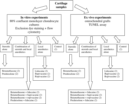
Methods: Cell viability and apoptosis/necrosis assessment of human articular chondrocytes were performed in vitro (chondrocyte cell cultures) and ex vivo (osteochondral specimens) using flow cytometry and TUNEL analysis, respectively.

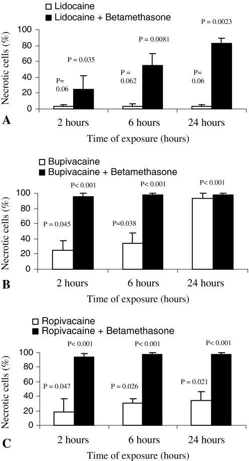
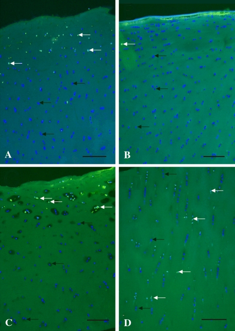
Results: Glucocorticoids and local anesthetics induce apoptosis in chondrocytes at various rates. When used in combination, the percentage of dead chondrocytes was increased in in vitro chondrocyte cell cultures and osteochondral ex vivo specimens.

Conclusions: We observed a time-dependent decrease in chondrocyte viability after concurrent steroid and local anesthetic exposure.

Clinical relevance: The combination of glucocorticoids and local anesthetics has an adverse effect on articular chondrocytes, and it raises a question regarding whether concomitant administration should be used in treating osteoarthritis.


Figures

Fig. 1
Fig. 1
A diagram of the experimental design is shown. All experiments were repeated three times.
Fig. 2
Fig. 2
An illustration of the osteochondral explants used for the ex vivo experiments is shown. The peripheral 2 mm from each side of the specimen was removed after the treatment because diffusion through the side cuts of the cartilage may bias the results. Only the remaining central portion of the cartilage was used for assessment.
Fig. 3A–F
Fig. 3A–F
The results of flow cytometry analysis of chondrocyte cell cultures treated with (A) control phosphate-buffered saline, (B) lidocaine, (C) betamethasone, (D) prednisolone, (E) betamethasone-lidocaine combination, and (F) prednisolone-lidocaine combination are shown. The time course changes of apoptotic and necrotic cells up to 24 hours of exposure with respect to the three detection points of 2, 6, and 24 hours are indicated in the X axis. The ratio of the apoptotic or necrotic cells is shown in the Y axis in percent. The bars represent the mean of three independent experiments with standard error as error bars. Results from the various experimental groups were compared with those of the PBS control group. Exact probability values also are shown as p values.
Fig. 4A–C
Fig. 4A–C
The ratios of necrotic chondrocytes treated with different local anesthetics alone or in combination with betamethasone are shown. Substantial differences are detectable between administration of (A) lidocaine or lidocaine + betamethasone, (B) bupivacaine or bupivacaine + betamethasone, and (C) ropivacaine or ropivacaine + betamethasone combinations. The X axis represents the exposure time in hours. The Y axis shows the ratio of necrotic chondrocytes in percent. The bars represent the mean of the three independent experiments with standard error as error bars. Results from the various experimental groups were compared with those of the PBS control group. Exact probability values are shown as p values.
Fig. 5A–D
Fig. 5A–D
An histologic assessment of the distribution of chondrocytes in articular cartilage explants was performed. The TUNEL assay (FITC-Annexin-V) with DAPI counterstaining shows apoptotic cells after 24 hours of treatment in osteochondral ex vivo specimens. The results of (A) lidocaine, (B) betamethasone, (C) prednisolone, and (D) prednisolone-lidocaine combinations are shown. The blue staining shows all nuclei of living cells (black arrows), illuminating green (TUNEL-positive) cells are dead (white arrows) (Original magnification, ×50). Scale bar = 150 μm.

References

    1. American College of Rheumatology Subcommittee on Osteoarthritis Guidelines Recommendations for the medical management of osteoarthritis of the hip and knee: 2000 update. Arthritis Rheum. 2000;43:1905–1915. doi: 10.1002/1529-0131(200009)43:9<1905::AID-ANR1>3.0.CO;2-P. - DOI - PubMed
    1. Arroll B, Goodyear-Smith F. Corticosteroid injections for osteoarthritis of the knee: meta-analysis. BMJ. 2004;328:869. doi: 10.1136/bmj.38039.573970.7C. - DOI - PMC - PubMed
    1. Bellamy N, Campbell J, Robinson V, Gee T, Bourne R, Wells G. Intraarticular corticosteroid for treatment of osteoarthritis of the knee. Cochrane Database Syst Rev. 2006;CD005328. - PubMed
    1. Bjordal JM, Klovning A, Ljunggren AE, Slordal L. Short-term efficacy of pharmacotherapeutic interventions in osteoarthritic knee pain: a meta-analysis of randomised placebo-controlled trials. Eur J Pain. 2007;11:125–138. doi: 10.1016/j.ejpain.2006.02.013. - DOI - PubMed
    1. Blanco FJ, Guitian R, Vazquez-Martul E, Toro FJ, Galdo F. Osteoarthritis chondrocytes die by apoptosis: a possible pathway for osteoarthritis pathology. Arthritis Rheum. 1998;41:284–289. doi: 10.1002/1529-0131(199802)41:2<284::AID-ART12>3.0.CO;2-T. - DOI - PubMed

LinkOut - more resources