

Local RAS
- PMID: 20700838
- PMCID: PMC7120160
- DOI: 10.1007/978-90-481-9060-7_5
Local RAS
Abstract
The concept of a circulating RAS is well established and known to play an endocrine role in the regulation of fluid homeostasis (see Section 4.1, Chapter 4). However, it is more appropriate to view the RAS in the contemporary notion as an “angiotensin-generating system”, which consists of angiotensinogen, angiotensin-generating enzymes, and angiotensins, as well as their receptors. Some RASs can be termed as “complete”, having renin and ACE involved in the biosynthesis of angiotensin II peptide, i.e. in a renin and/or ACE-dependent manner which is exemplified in the circulating RAS. On the other hand, some RAS can be termed as “partial”, having alternate enzymes to renin and ACE, such as chymase and ACE2 (see Section 4.3, Chapter 4) available for the generation of angiotensin II and other bioactive angiotensin peptides in the biosynthetic cascade, i.e. in a renin and/or ACE-independent manner. Complete vs. partial RASs can be exemplified in the so-called intrinsic angiotensin-generating system or local RAS; for example, a local and functional RAS with renin and ACE-dependent but a renin-independent pathway have been indentified in the pancreas and carotid body, respectively. In the past two decades, local RASs have gained increasing recognition especially with regards to their clinical importance. Distinct from the circulating RAS, these functional local RASs exist in such diverse tissues and organs as the pancreas, liver, intestine, heart, kidney, vasculature, carotid body, and adipose, as well as the nervous, reproductive, and digestive systems. Taken into previous findings from our laboratory and others together, Table 5.1 is a summary of some recently identified local RASs in various levels of tissues and organs.
Figures

Similar articles
-
Novel roles of a local angiotensin-generating system in the carotid body.J Physiol. 2006 Aug 15;575(Pt 1):4. doi: 10.1113/jphysiol.2006.115550. Epub 2006 Jun 29. J Physiol. 2006. PMID: 16809356 Free PMC article. No abstract available.
-
Renin-angiotensin system in the carotid body.Int J Biochem Cell Biol. 2003 Jun;35(6):847-54. doi: 10.1016/s1357-2725(02)00180-2. Int J Biochem Cell Biol. 2003. PMID: 12676171 Review.
-
A locally generated angiotensin system in rat carotid body.Regul Pept. 2002 Jul 15;107(1-3):97-103. doi: 10.1016/s0167-0115(02)00068-x. Regul Pept. 2002. PMID: 12137971
-
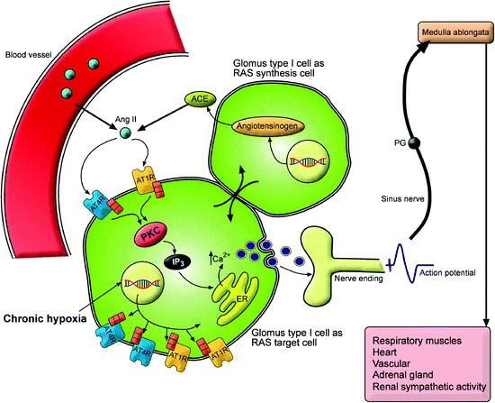
Upregulation of a local renin-angiotensin system in the rat carotid body during chronic intermittent hypoxia.Exp Physiol. 2014 Jan;99(1):220-31. doi: 10.1113/expphysiol.2013.074591. Epub 2013 Sep 13. Exp Physiol. 2014. PMID: 24036592
-
LOCAL RENIN-ANGIOTENSIN SYSTEM OF SMALL INTESTINE.Eksp Klin Gastroenterol. 2016 Jul;12(12):97-104. Eksp Klin Gastroenterol. 2016. PMID: 29889431 Review. English, Russian.
Cited by
-
The Impact of ACE and ACE2 Gene Polymorphisms in Pulmonary Diseases Including COVID-19.In Vivo. 2022 Jan-Feb;36(1):13-29. doi: 10.21873/invivo.12672. In Vivo. 2022. PMID: 34972696 Free PMC article. Review.
-
Novel Insights into the Antagonistic Effects of Losartan against Angiotensin II/AGTR1 Signaling in Glioblastoma Cells.Cancers (Basel). 2021 Sep 10;13(18):4555. doi: 10.3390/cancers13184555. Cancers (Basel). 2021. PMID: 34572782 Free PMC article.
-
Increased Endogenous Activity of the Renin-Angiotensin System Reduces Infarct Size in the Rats with Early Angiotensin II-dependent Hypertension which Survive the Acute Ischemia/Reperfusion Injury.Front Pharmacol. 2021 May 28;12:679060. doi: 10.3389/fphar.2021.679060. eCollection 2021. Front Pharmacol. 2021. PMID: 34122103 Free PMC article.
-
The Renin-Angiotensin System in Liver Disease.Int J Mol Sci. 2024 May 27;25(11):5807. doi: 10.3390/ijms25115807. Int J Mol Sci. 2024. PMID: 38891995 Free PMC article. Review.
-
Inflammation Triggered by SARS-CoV-2 and ACE2 Augment Drives Multiple Organ Failure of Severe COVID-19: Molecular Mechanisms and Implications.Inflammation. 2021 Feb;44(1):13-34. doi: 10.1007/s10753-020-01337-3. Epub 2020 Oct 8. Inflammation. 2021. PMID: 33029758 Free PMC article. Review.
References
-
- Alison MR. Regulation of hepatic growth. Physiol Rev. 1986;66:499–451. - PubMed
-
- Arroyo V, Bosch J, Mauri M, Ribera F, Navarro-Lopez F, Rodes J. Effect of angiotensin-II blockade on systemic and hepatic haemodynamics and on the renin-angiotensin-aldosterone system in cirrhosis with ascites. Eur J Clin Invest. 1981;11:221–229. doi: 10.1111/j.1365-2362.1981.tb01844.x. - DOI - PubMed
Publication types
MeSH terms
LinkOut - more resources
Full Text Sources
Miscellaneous
