. 2010 Oct 15;431(2):311-20.
doi: 10.1042/BJ20101100.

Contraction regulates site-specific phosphorylation of TBC1D1 in skeletal muscle

Affiliations

Contraction regulates site-specific phosphorylation of TBC1D1 in skeletal muscle

Kanokwan Vichaiwong et al. Biochem J. .

Abstract

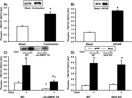
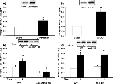
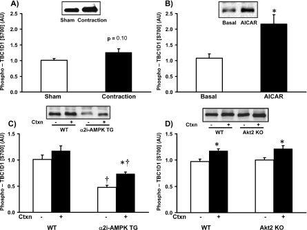
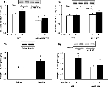
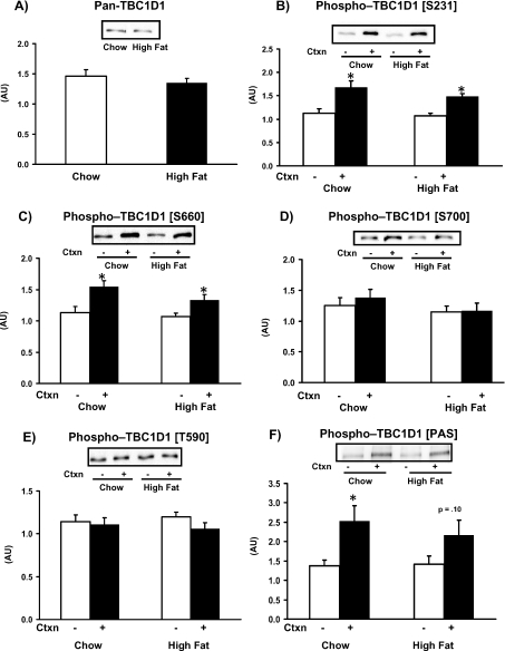
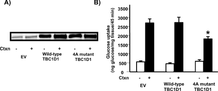
TBC1D1 (tre-2/USP6, BUB2, cdc16 domain family member 1) is a Rab-GAP (GTPase-activating protein) that is highly expressed in skeletal muscle, but little is known about TBC1D1 regulation and function. We studied TBC1D1 phosphorylation on three predicted AMPK (AMP-activated protein kinase) phosphorylation sites (Ser231, Ser660 and Ser700) and one predicted Akt phosphorylation site (Thr590) in control mice, AMPKα2 inactive transgenic mice (AMPKα2i TG) and Akt2-knockout mice (Akt2 KO). Muscle contraction significantly increased TBC1D1 phosphorylation on Ser231 and Ser660, tended to increase Ser700 phosphorylation, but had no effect on Thr590. AICAR (5-aminoimidazole-4-carboxyamide ribonucleoside) also increased phosphorylation on Ser231, Ser660 and Ser700, but not Thr590, whereas insulin only increased Thr590 phosphorylation. Basal and contraction-stimulated TBC1D1 Ser231, Ser660 and Ser700 phosphorylation were greatly reduced in AMPKα2i TG mice, although contraction still elicited a small increase in phosphorylation. Akt2 KO mice had blunted insulin-stimulated TBC1D1 Thr590 phosphorylation. Contraction-stimulated TBC1D1 Ser231 and Ser660 phosphorylation were normal in high-fat-fed mice. Glucose uptake in vivo was significantly decreased in tibialis anterior muscles overexpressing TBC1D1 mutated on four predicted AMPK phosphorylation sites. In conclusion, contraction causes site-specific phosphorylation of TBC1D1 in skeletal muscle, and TBC1D1 phosphorylation on AMPK sites regulates contraction-stimulated glucose uptake. AMPK and Akt regulate TBC1D1 phosphorylation, but there must be additional upstream kinases that mediate TBC1D1 phosphorylation in skeletal muscle.


Figures

Figure 1
Figure 1. TBC1D1 Ser231 phosphorylation is increased by contraction and AICAR
(A) Contraction (Ctxn) increased TBC1D1 Ser231 phosphorylation in tibialis anterior muscles from ICR mice. Mice were anaesthetized and contraction was generated by electrical stimulation of the peroneal nerve for 15 min, while the contra-lateral side served as a sham-operated control. (B) AICAR increased TBC1D1 Ser231 phosphorylation. EDL muscles were isolated from ICR mice and incubated with 2 mM AICAR or vehicle for 40 min. (C) Phosphorylation of TBC1D1 Ser231 was reduced in AMPKα2i TG mice. AMPKα2i TG mice and WT littermates underwent contraction as described in (A). (D) Basal and contraction-stimulated TBC1D1 Ser231 was preserved in Akt2 KO mice. Akt2 KO mice and WT littermates underwent contraction as described in (A). Data are means±S.E.M.; n = 5–7/group. *P< 0.05 compared with control; †P< 0.05 compared with corresponding WT. AU, arbitrary units.
Figure 2
Figure 2. TBC1D1 Ser660 phosphorylation is regulated by contraction and AICAR
Experimental details are as described in the legend to Figure 1. (A) Contraction (Ctxn) in situ for 15 min increased TBC1D1 Ser660 phosphorylation in tibialis anterior muscles from ICR mice. (B) AICAR increased TBC1D1 Ser660 phosphorylation. (C) Phosphorylation of TBC1D1 Ser660 was reduced in AMPKα2i TG mice. (D) Basal and contraction-stimulated TBC1D1 Ser660 was preserved in Akt2 KO mice. Data are means±S.E.M.; n = 5–7/group. *P< 0.05 compared with control; †P< 0.05 compared with corresponding WT. AU, arbitrary units.
Figure 3
Figure 3. TBC1D1 Ser700 phosphorylation is increased by AICAR, but only responds minimally to contraction
Experimental details are as described in the legend to Figure 1. (A) Contraction (Ctxn) in situ for 15 min tended to increase TBC1D1 Ser700 phosphorylation in tibialis anterior muscles from ICR mice. (B) AICAR increased TBC1D1 Ser700 phosphorylation. (C) Phosphorylation of TBC1D1 Ser700 was reduced in AMPKα2i TG mice. (D) TBC1D1 Ser700 phosphorylation was preserved in Akt2 KO mice. Data are means±S.E.M.; n = 5–7/group. *P< 0.05 compared with control; †P< 0.05 compared with corresponding WT. AU, arbitrary units.
Figure 4
Figure 4. TBC1D1 Thr590 phosphorylation is regulated by insulin, but not contraction or AICAR
(A) Phosphorylation of TBC1D1 Ser590 was significantly reduced in AMPKα2i TG mice at rest. Contraction (Ctxn) in situ for 15 min increased phosphorylation in AMPKα2i TG mice, but the increase after contraction in WT mice was not statistically significant. (B) The response to contraction was not affected in Akt2 KO mice. (C) Maximal insulin injection increased TBC1D1 Ser590 phosphorylation in ICR mice. Mice were fasted and injected intraperitoneally with maximal insulin or saline, and tibialis anterior muscles were dissected 10 min later. (D) Maximal insulin injection increased TBC1D1 Ser590 phosphorylation in WT mice, and the effect was ablated in Akt2 KO mice. Treatment was the same as described in (C). Data are means±S.E.M.; n = 5–7/group. *P< 0.05 compared with control; †P< 0.05 compared with corresponding WT. AU, arbitrary units.
Figure 5
Figure 5. High-fat diet does not affect contraction-stimulated TBC1D1 phosphorylation
(A) TBC1D1 expression was not altered in tibialis anterior muscles of mice fed on a high-fat diet (60% kcal of fat) for 9 weeks. (BF) High-fat feeding had no effect on basal or contraction-stimulated levels of (B) TBC1D1 Ser231 phosphorylation, (C) TBC1D1 Ser660 phosphorylation, (D) TBC1D1 Ser700 phosphorylation, (E) TBC1D1 Thr590 or (F) PAS phosphorylation at 150–160 kDa. Data are means±S.E.M.; n = 6/group. *P< 0.05 compared with control. AU, arbitrary units; Ctxn, contraction.
Figure 6
Figure 6. Overexpression of the TBC1D1 4A mutant impairs contraction-stimulated glucose uptake
To determine whether TBC1D1 phosphorylation at AMPK consensus sites regulates glucose uptake in contracted muscle, four phosphorylation sites on TBC1D1 (Ser231, Thr499, Ser660 and Ser700) were mutated to alanine (4A) and this mutant and WT TBC1D1 were expressed in tibialis anterior muscles. (A) Representative Western blot of total TBC1D1 showing overexpression 7 days after plasmid DNA injections. (B) Contraction-stimulated glucose uptake was not affected by overexpression of WT TBC1D1, whereas expression of the TBC1D1 4A mutant impaired contraction-stimulated glucose uptake by 42%. Data are means±S.E.M.; n = 8–15/group. *P< 0.05 compared with contraction-stimulated empty vector (EV) controls. Ctxn, contraction.

References

    1. DeFronzo R. A., Jacot E., Jequier E., Maeder E., Wahren J., Felber J. P. The effect of insulin on the disposal of intravenous glucoseResults from indirect calorimetry and hepatic and femoral venous catheterization. Diabetes. 1981;30:1000–1007. - PubMed
    1. Baron A. D., Brechtel G., Wallace P., Edelman S. V. Rates and tissue sites of non-insulin- and insulin-mediated glucose uptake in humans. Am. J. Physiol. 1988;255:E769–E774. - PubMed
    1. DeFronzo R. A., Simonson D., Ferrannini E. Hepatic and peripheral insulin resistance: a common feature of type II (non-insulin-dependent) and type I (insulin dependent) diabetes mellitus. Diabetologia. 1982;23:313–319. - PubMed
    1. Dohm G. L., Tapscott E. B., Pories W. J., Dabbs D. J., Flickinger E. G., Meelheim D., Fushiki T., Atkinson S. M., Elton C. W., Caro J. F. An in vitro human muscle preparation suitable for metabolic studies. Decreased insulin stimulation of glucose transport in muscle from morbidly obese and diabetic subjects. J. Clin. Invest. 1988;82:486–494. - PMC - PubMed
    1. Kennedy J. W., Hirshman M. F., Gervino E. V., Ocel J. V., Forse R. A., Hoenig S. J., Aronson D., Goodyear L. J., Horton E. S. Acute exercise induces GLUT4 translocation in skeletal muscle of normal human subjects and subjects with type 2 diabetes. Diabetes. 1999;48:1192–1197. - PubMed

Publication types

MeSH terms