Review
. 2010 Oct;9(5):667-84.
doi: 10.1111/j.1474-9726.2010.00608.x. Epub 2010 Aug 15.

Fat tissue, aging, and cellular senescence

Affiliations
Free PMC article
Review

Fat tissue, aging, and cellular senescence

Tamara Tchkonia et al. Aging Cell. 2010 Oct.
Free PMC article

Abstract

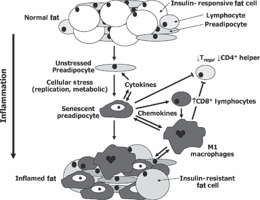
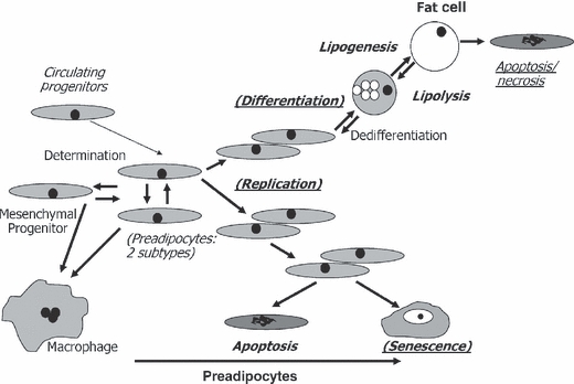
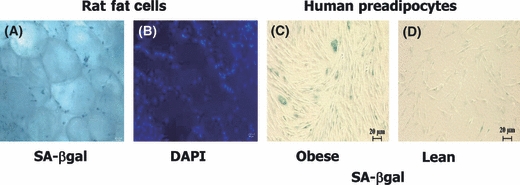
Fat tissue, frequently the largest organ in humans, is at the nexus of mechanisms involved in longevity and age-related metabolic dysfunction. Fat distribution and function change dramatically throughout life. Obesity is associated with accelerated onset of diseases common in old age, while fat ablation and certain mutations affecting fat increase life span. Fat cells turn over throughout the life span. Fat cell progenitors, preadipocytes, are abundant, closely related to macrophages, and dysdifferentiate in old age, switching into a pro-inflammatory, tissue-remodeling, senescent-like state. Other mesenchymal progenitors also can acquire a pro-inflammatory, adipocyte-like phenotype with aging. We propose a hypothetical model in which cellular stress and preadipocyte overutilization with aging induce cellular senescence, leading to impaired adipogenesis, failure to sequester lipotoxic fatty acids, inflammatory cytokine and chemokine generation, and innate and adaptive immune response activation. These pro-inflammatory processes may amplify each other and have systemic consequences. This model is consistent with recent concepts about cellular senescence as a stress-responsive, adaptive phenotype that develops through multiple stages, including major metabolic and secretory readjustments, which can spread from cell to cell and can occur at any point during life. Senescence could be an alternative cell fate that develops in response to injury or metabolic dysfunction and might occur in nondividing as well as dividing cells. Consistent with this, a senescent-like state can develop in preadipocytes and fat cells from young obese individuals. Senescent, pro-inflammatory cells in fat could have profound clinical consequences because of the large size of the fat organ and its central metabolic role.


Figures

Fig. 1
Fig. 1
Hypothetical model of the chain of events culminating in fat tissue inflammation in obesity. Preadipocytes and fat tissue endothelial cells may acquire an activated, pro-inflammatory, senescent-like phenotype in response to repeated replication, fatty acids, toxic metabolites, chronically high IGF-1, glucose, or other stimuli (gray = inflamed). Inflammatory cytokines could spread this activated secretory phenotype from cell to cell and block full differentiation of preadipocytes into insulin-responsive fat cells, amplifying the process. Chemokines, cytokines, and ECM modifiers produced by pro-inflammatory cells might activate adaptive immune responses. Shifts from anti- to proinflammatory T-lymphocyte subsets and mast cell infiltration owing to cytokine and lymphokine production, toxic metabolites (including fatty acids and reactive oxygen species), and cytokines released by inflamed preadipocytes and endothelial cells may combine to promote M1 macrophage activation. The inflammatory cytokines could induce systemic effects, further impede adipogenesis, and promote fat cell lipolysis, releasing fatty acids that aggravate the fat tissue pro-inflammatory state and cause systemic lipotoxicity. Similar processes could be involved in age-related fat tissue dysregulation and metabolic dysfunction. Some of these processes appear to vary in extent among fat depots in obesity (Feuerer et al., 2009; Nishimura et al., 2009; Winer et al., 2009) as well as aging (Cartwright et al., 2010).
Fig. 2
Fig. 2
Impact of aging, obesity, anatomic origin, and serial passage on cell dynamic mechanisms of fat tissue turnover. Up to 50% of cells in fat tissue are committed preadipocytes that arise from multipotent, slowly replicating mesenchymal progenitor cells and possibly circulating progenitors (Hong et al., 2005; Crossno et al., 2006). Preadipocyte numbers are maintained by replication. Preadipocytes can reversibly switch into a slowly replicating subtype, can become macrophage-like, and may be able to progress up or down the adipocytic lineage (Cousin et al., 1999; Charriere et al., 2003; Tchkonia et al., 2005; Gustafson et al., 2009). Preadipocytes are depleted by differentiation into fat cells, apoptosis, necrosis, and cellular senescence. Enlargement of fat cells (lipid accumulation) and maintenance of insulin responsiveness are tied to processes initiated during differentiation, including adipogenic transcription factor expression. Fat cells, especially large fat cells, can be removed by a process with features of both apoptosis and necrosis and that can induce inflammation. The balance among these cell dynamic properties determines preadipocyte and fat cell numbers. Cell dynamic processes that vary with aging are in bold, obesity are underlined, anatomic origin in Italics, and serial passage in parentheses. These differences persist for at least 40 population doublings in cloned preadipocytes in the case of anatomic origin, 16 in aging, and 8 in obesity.
Fig. 3
Fig. 3
Senescent preadipocytes can accumulate in fat tissue of even young individuals. Freshly isolated, whole perirenal fat tissue isolated from 2-month-old obese male Zucker rats was assayed for senescence-associated β-galactosidase (A; SA β-gal) or stained with DAPI to show nuclei (B; representative of N = 3 animals). Preadipocytes cultured from young obese rats had senescent-associated heterochromatic foci. Senescent cells are also increased in high fat-fed mice, express p53 (Minamino et al., 2009), and are less frequent in age-matched ad libitum fed controls. Human preadipocytes from an obese young adult subject were SA β-gal positive (C; age 21 years.; body mass index [BMI] 50; representative of six obese subjects), while fewer preadipocytes cultured from a lean subject were senescent (D; age 26 years.; BMI 23; representative of nine lean subjects). Nevertheless, occasional senescent cells were found in preadipocytes cultured from all nine young, lean subjects.

Similar articles

Cited by

References

    1. Abel ED, Peroni O, Kim JK, Kim YB, Boss O, Hadro E, Minnemann T, Shulman GI, Kahn BB. Adipose-selective targeting of the GLUT4 gene impairs insulin action in muscle and liver. Nature. 2001;409:729–733. - PubMed
    1. Ahima RS. Connecting obesity, aging and diabetes. Nat. Med. 2009;15:996–997. - PubMed
    1. Argmann C, Dobrin R, Heikkinen S, Auburtin A, Pouilly L, Cock TA, Koutnikova H, Zhu J, Schadt EE, Auwerx J. Ppargamma2 is a key driver of longevity in the mouse. PLoS Genet. 2009;5:e1000752. - PMC - PubMed
    1. Atzmon G, Pollin TI, Crandall J, Tanner K, Schechter CB, Scherer PE, Rincon M, Siegel G, Katz M, Lipton RB, Shuldiner AR, Barzilai N. Adiponectin levels and genotype: a potential regulator of life span in humans. J. Gerontol. A Biol. Sci. Med. Sci. 2008;63:447–453. - PMC - PubMed
    1. Baker DJ, Jeganathan KB, Malureanu L, Perez-Terzic C, Terzic A, van Deursen JM. Early aging-associated phenotypes in Bub3/Rae1 haploinsufficient mice. J. Cell Biol. 2006;172:529–540. - PMC - PubMed

Publication types