

Iron sensitizes keratinocytes and fibroblasts to UVA-mediated matrix metalloproteinase-1 through TNF-α and ERK activation
- PMID: 20701626
- PMCID: PMC3022993
- DOI: 10.1111/j.1600-0625.2010.01152.x
Iron sensitizes keratinocytes and fibroblasts to UVA-mediated matrix metalloproteinase-1 through TNF-α and ERK activation
Abstract
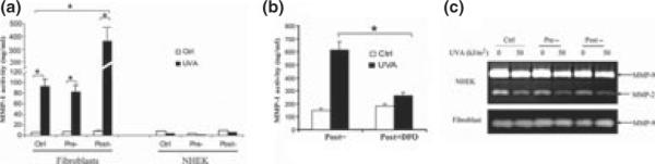
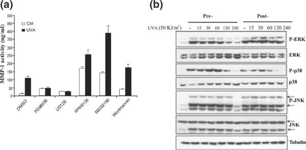
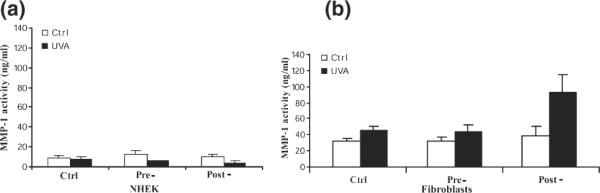
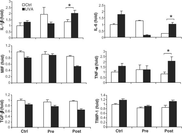
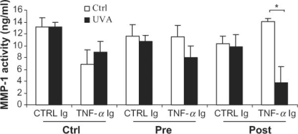
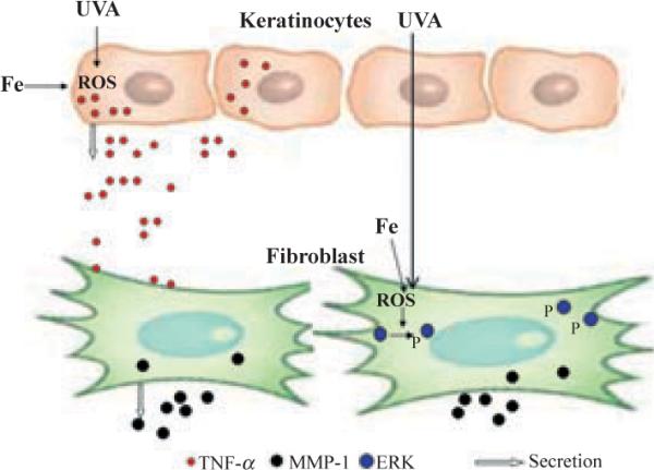
Oestrogen deficiency is regarded as the main causative factor in postmenopausal skin ageing and photoageing. While women after menopause experience low levels of oestrogen because of cease of ovarian function, they are also exposed to high levels of iron as a result of cessation of menstruation. In this study, we investigated whether this increase in iron presents a risk to the postmenopausal skin. Because of the lack of appropriate animal models to closely mimic the low oestrogen and high iron conditions, we tested the hypothesis in a high iron and low oestrogen culture model. Here, we showed that primary human dermal fibroblasts exposed to iron did not affect the baseline levels of matrix metalloproteinase-1 (MMP-1) activity. However, the iron-exposed fibroblasts were sensitized to UVA exposure, which resulted in a synergistic increase in MMP-1. UVA activated the three members of MAPK family: ERKs, p38, and JNKs. Additional activation of ERKs by iron contributed to the synergistic increases. Primary normal human epidermal keratinocytes (NHEK) did not respond to iron or UVA exposure as measured by MMP-1, but produced tumor necrosis factor-alpha (TNF-α) in the media, which then stimulated MMP-1 in fibroblasts. Our results indicate that iron and UVA increase MMP-1 activity in dermal fibroblasts not only directly through ERK activation but also by an indirect paracrine loop through TNF-α released by NHEK. We conclude that in addition to oestrogen deficiency, increased iron as a result of menopause could be a novel risk factor by sensitizing postmenopausal skin to solar irradiation.
© 2010 John Wiley & Sons A/S.
Figures
Similar articles
-
IL-1 receptor antagonist attenuates MAP kinase/AP-1 activation and MMP1 expression in UVA-irradiated human fibroblasts induced by culture medium from UVB-irradiated human skin keratinocytes.Int J Mol Med. 2005 Dec;16(6):1117-24. Int J Mol Med. 2005. PMID: 16273295
-
Syringaresinol Inhibits UVA-Induced MMP-1 Expression by Suppression of MAPK/AP-1 Signaling in HaCaT Keratinocytes and Human Dermal Fibroblasts.Int J Mol Sci. 2020 Jun 1;21(11):3981. doi: 10.3390/ijms21113981. Int J Mol Sci. 2020. PMID: 32492931 Free PMC article.
-
IL-17 stimulates MMP-1 expression in primary human cardiac fibroblasts via p38 MAPK- and ERK1/2-dependent C/EBP-beta , NF-kappaB, and AP-1 activation.Am J Physiol Heart Circ Physiol. 2007 Dec;293(6):H3356-65. doi: 10.1152/ajpheart.00928.2007. Epub 2007 Oct 5. Am J Physiol Heart Circ Physiol. 2007. PMID: 17921324
-
Predominant activation of MAP kinases and pro-destructive/pro-inflammatory features by TNF alpha in early-passage synovial fibroblasts via TNF receptor-1: failure of p38 inhibition to suppress matrix metalloproteinase-1 in rheumatoid arthritis.Ann Rheum Dis. 2007 Aug;66(8):1043-51. doi: 10.1136/ard.2006.062521. Epub 2007 Jan 12. Ann Rheum Dis. 2007. PMID: 17223661 Free PMC article.
-
TNF-alpha production in the skin.Arch Dermatol Res. 2009 Jan;301(1):87-91. doi: 10.1007/s00403-008-0893-7. Epub 2008 Sep 30. Arch Dermatol Res. 2009. PMID: 18825399 Review.
Cited by
-
Shedding a New Light on Skin Aging, Iron- and Redox-Homeostasis and Emerging Natural Antioxidants.Antioxidants (Basel). 2022 Feb 27;11(3):471. doi: 10.3390/antiox11030471. Antioxidants (Basel). 2022. PMID: 35326121 Free PMC article. Review.
-
Photoprotection by dietary phenolics against melanogenesis induced by UVA through Nrf2-dependent antioxidant responses.Redox Biol. 2016 Aug;8:79-90. doi: 10.1016/j.redox.2015.12.006. Epub 2015 Dec 19. Redox Biol. 2016. PMID: 26765101 Free PMC article.
-
Transdermal Delivery of Functional Collagen Via Polyvinylpyrrolidone Microneedles.Ann Biomed Eng. 2015 Dec;43(12):2978-90. doi: 10.1007/s10439-015-1353-0. Epub 2015 Jun 12. Ann Biomed Eng. 2015. PMID: 26066056 Free PMC article.
-
Evaluation of Antioxidant and Anti-Inflammatory Effects of a Nanoformulation Derived from Annurca Apple Callus Extract in an In Vitro Model of Iron Overload-Induced Inflammation.Antioxidants (Basel). 2025 May 24;14(6):631. doi: 10.3390/antiox14060631. Antioxidants (Basel). 2025. PMID: 40563266 Free PMC article.
-
Aberrant Transferrin and Ferritin Upregulation Elicits Iron Accumulation and Oxidative Inflammaging Causing Ferroptosis and Undermines Estradiol Biosynthesis in Aging Rat Ovaries by Upregulating NF-Κb-Activated Inducible Nitric Oxide Synthase: First Demonstration of an Intricate Mechanism.Int J Mol Sci. 2022 Oct 21;23(20):12689. doi: 10.3390/ijms232012689. Int J Mol Sci. 2022. PMID: 36293552 Free PMC article.
References
-
- Elias PM, Ghadially R. Clin Geriatr Med. 2002;18:103–120. - PubMed
-
- Helfrich YR, Sachs DL, Voorhees JJ. Dermatol Nurs. 2008;20:177–183. quiz 184. - PubMed
-
- Rabe JH, Mamelak AJ, McElgunn PJ, et al. J Am Acad Dermatol. 2006;55:1–19. - PubMed
-
- Yaar M, Gilchrest BA. J Investig Dermatol Symp Proc. 1998;3:47–51. - PubMed
-
- El-Domyati M, Attia S, Saleh F, et al. Exp Dermatol. 2002;11:398–405. - PubMed
Publication types
MeSH terms
Substances
Grants and funding
LinkOut - more resources
Full Text Sources
Medical
Miscellaneous
