. 2010 Aug 11:11:64.
doi: 10.1186/1471-2121-11-64.

HMGA1 down-regulation is crucial for chromatin composition and a gene expression profile permitting myogenic differentiation

Affiliations

HMGA1 down-regulation is crucial for chromatin composition and a gene expression profile permitting myogenic differentiation

Jan Brocher et al. BMC Cell Biol. .

Abstract

Background: High mobility group A (HMGA) proteins regulate gene transcription through architectural modulation of chromatin and the formation of multi-protein complexes on promoter/enhancer regions. Differential expression of HMGA variants has been found to be important for distinct differentiation processes and deregulated expression was linked to several disorders. Here we used mouse C2C12 myoblasts and C2C12 cells stably over-expressing HMGA1a-eGFP to study the impact of deregulated HMGA1 expression levels on cellular differentiation.

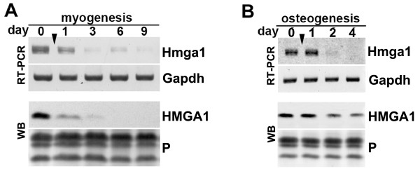
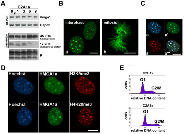
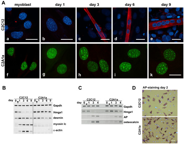
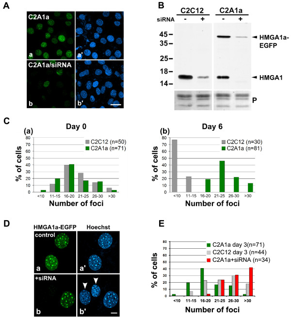
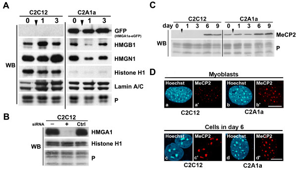
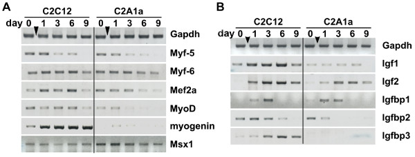
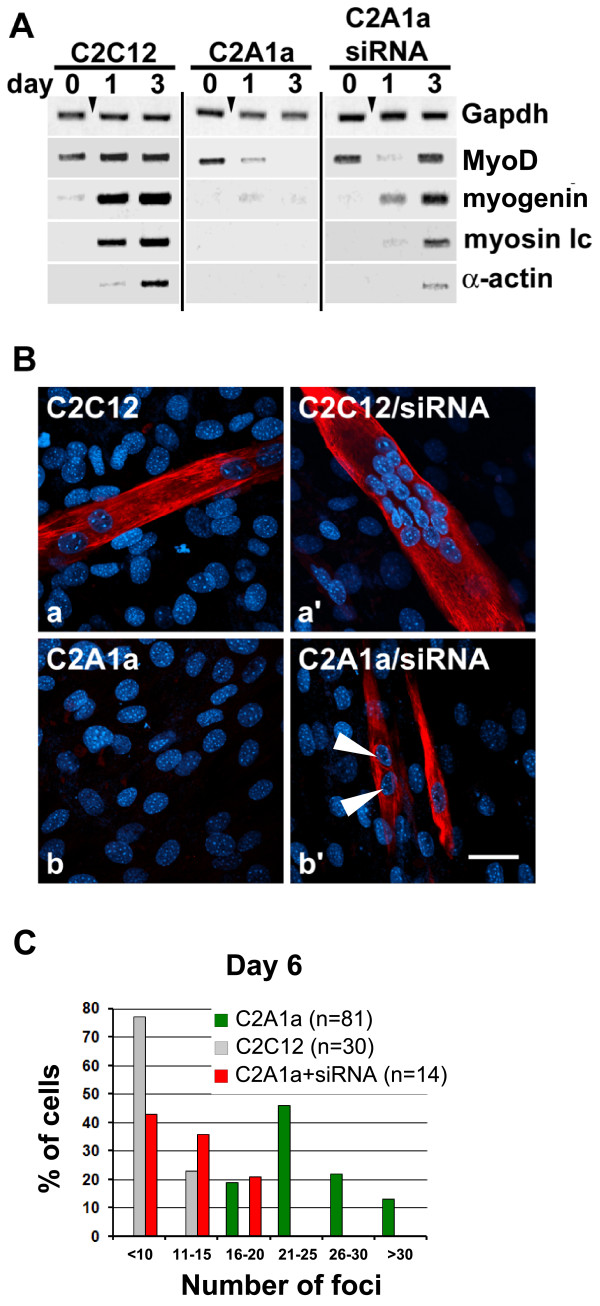
Results: We found that induction of the myogenic or osteogenic program of C2C12 cells caused an immediate down-regulation of HMGA1. In contrast to wild type C2C12 cells, an engineered cell line with stable over-expression of HMGA1a-eGFP failed to differentiate into myotubes. Immunolocalization studies demonstrated that sustained HMGA1a-eGFP expression prevented myotube formation and chromatin reorganization that normally accompanies differentiation. Western Blot analyses showed that elevated HMGA1a-eGFP levels affected chromatin composition through either down-regulation of histone H1 or premature expression of MeCP2. RT-PCR analyses further revealed that sustained HMGA1a expression also affected myogenic gene expression and caused either down-regulation of genes such as MyoD, myogenin, Igf1, Igf2, Igfbp1-3 or up-regulation of the transcriptional repressor Msx1. Interestingly, siRNA experiments demonstrated that knock-down of HMGA1a was required and sufficient to reactivate the myogenic program in induced HMGA1a over-expressing cells.

Conclusions: Our data demonstrate that HMGA1 down-regulation after induction is required to initiate the myogenic program in C2C12 cells. Sustained HMGA1a expression after induction prevents expression of key myogenic factors. This may be due to specific gene regulation and/or global effects on chromatin. Our data further corroborate that altered HMGA1 levels influence the expression of other chromatin proteins. Thus, HMGA1 is able to establish a specific chromatin composition. This work contributes to the understanding of how differential HMGA1 expression is involved in chromatin organization during cellular differentiation processes and it may help to comprehend effects of HMGA1 over-expression occurring in malign or benign tumours.


Figures

Figure 1
Figure 1
Differential expression of HMGA1 proteins during C2C12 cell differentiation. (A) Down-regulation of HMGA1 during myogenic differentiation of C2C12 cells as analyzed by PCR and Western blotting (WB). For Western blots proteins of 1.5 × 10⁵ nuclei were separated on a 15% SDS-polyacrylamide gel. For RT-PCR 1 μg of total RNA was used to produce cDNAs for the PCR reaction. For PCR identical amounts of cDNA were used. Time points of analyses are indicated in days. The arrowhead marks the time point of induction. Day 0 denotes non-induced myoblasts. Gapdh expression served as a control for the reverse transcription in RT-PCR experiments. As a control for loading and Western blotting Ponceau staining (P) of core histones is shown. (B) Down-regulation of HMGA1 expression during osteogenesis. Osteogenesis in C2C12 cells was induced with 0.5 μg/ml BMP2 (arrowhead) and HMGA1 expression was analyzed at day 0 and on days 1, 3 and 4 after by RT-PCR (RT) and Western blot (WB) as described in (A).
Figure 2
Figure 2
Characterization of the HMGA1a-eGFP over-expressing cell line C2A1a. (A) Sustained expression of HMGA1a-eGFP in C2A1a cells throughout myogenic induction analyzed by RT-PCR and Western blotting (WB) as indicated. Both, fusion protein (~45 kDa) and endogenous protein (~17 kDa) were detected using an HMGA1-specific antibody. Note the sustained expression throughout myogenic induction. Loading controls are as mentioned in Fig. 1. (B) Localization of HMGA1a-eGFP in living C2A1a cells. Note the concentrated localization in heterochromatin foci (chromocenters) during interphase (a) and on pericentromeric regions during mitosis (b). Scale bars represent 10 μm. (C) Immunolocalization on fixed C2A1a cells using HP1α-specific antibodies. Note the colocalization of HMGA1a-eGFP (a') and HP1α (a'') in the chromocenters of C2A1a cells. DNA was stained with Hoechst (a). An overlay of a-a'' is shown in a'''. The bar represents 10 μm. (D) HMGA1a-eGFP (green) colocalizes with the H3K9me3 and H4K20me3 specific immunolocalizations (red) in C2A1a cells. DNA was stained with Hoechst. The bar represents 10 μm. (E) Cell cycle phases are unaffected in the C2A1a cell line. DNA from C2C12 and C2A1a was stained with propidium iodide and 20,000 cells from each cell line were analyzed by FACS. Cell numbers (counts) are plotted against the relative DNA content of the cells. Phase distribution was analyzed with modfit Lt3.1. 56.14% of C2C12 cells were in G1 phase, 29.54% in S phase, and 14.33% in G2 phase. In C2A1a cells 60.95% of the cells were in G1 phase, 24.43% in S phase, and 14.63% in G2 phase.
Figure 3
Figure 3
HMGA1a over-expression prevents myogenic differentiation. (A) Immunofluorescence localization of myosin (red) in C2C12 (a-e) and in C2A1a cells (f-k) before (myoblast) and after induction (days 1 to 9). All bars represent 20 μm. Myotube formation was only observed in wild type C2C12 cells. In pictures a-e DNA staining by Hoechst is shown in blue, respectively. HMGA1a-eGFP is shown in green. (B) RT-PCR analysis to compare the expression of the myosin light chain (myosin lc) and α-actin in C2C12 cells (left) and in C2A1a cells (right) as described in Fig. 1. Expression of Hmga1, Gapdh and desmin are shown as controls. (C) RT-PCR to analyze expression profiles of marker genes for osteogenesis. Genes analyzed were alkaline phosphatase (AP) and osteocalcin. Gapdh expression is shown as control. HMGA1a over-expression did not affect AP and osteocalcin transcription. (D) Alkaline phosphatase activity as marker for osteogenesis of C2C12 and C2A1a cells on day 2 of differentiation (bright field images). AP activity was visualized using NBT/BCIP staining. Shown are overlays of bright field images and fluorescence images with corresponding DNA staining. Scale bar is 50 μm.
Figure 4
Figure 4
HMGA1a over-expression interferes with chromatin organization during myogenesis. (A) Depletion of HMGA1a-eGFP after HMGA1a-siRNA treatment is visual through the loss of eGFP fluorescence in C2A1a cells (b). Mock transfected C2A1a cells are shown in a. The corresponding Hoechst stained images are shown in a' and b'. The bar represents 20 μm. (B) Western blot analysis showed considerable depletion of endogenous and eGFP-tagged HMGA1a proteins, respectively, 12-24 hours after siRNA treatment of C2C12 and C2A1a cells. Ponceau staining (P) of the core histones is presented as loading control. Protein molecular mass is indicated in kDa. (C) Heterochromatin foci (chromocenters) in C2C12 and C2A1a cells were quantified (n = cell number) and plotted as indicated. (a) In myoblasts (day 0) the number of chromocenters is identical in both cell lines. (b) During terminal muscle differentiation of the C2C12 cells, the number of heterochromatin foci decreases due to chromocenter clustering (grey columns) whereas the chromocenter number in C2A1a cells remains comparable to the number in myoblasts (green columns). (D) C2A1a control cells (a) and C2A1a cells 12-24 hours after siRNA treatment (b). HMGA1 depletion, indicated by absence of HMGA1a-eGFP, results in a higher number and a reduced size of chromocenters (arrowheads) (b'). The bar represents 10 μm. (E) Depletion of HMGA1 by siRNA in C2A1a myoblasts increased the chromocenter number (red columns) compared to C2A1a control cells (green columns). A similar chromocenter dissociation was detected in C2C12 wild type cells on differentiation day 3 (grey columns) prior to fusion during terminal differentiation.
Figure 5
Figure 5
HMGA1a over-expression alters the chromatin composition. (A) Western blot analyses comparing the expression of HMGB1, HMGN1, and histone H1 in C2C12 cells and C2A1a cells in myoblasts (day 0) and on days 1 and 3 of differentiation. The antibody used [43] recognizes all histone H1 variants. Proteins of 1.5 × 10⁵ cells were analyzed in 15% SDS-PAGE. Lamin A/C expression and Ponceau S staining are shown as loading controls. (B) Histone H1 levels are unaffected in C2C12 cells after HMGA1 knock-down. Western blot was as described in Fig. 5A. Antibodies used are indicated. Histone H1 antibody used was directed against all H1-sub-variants (Abcam). Cells were untreated (-), siRNA treated (+) or transfected using siRNA control oligos (Ctrl). Ponceau S staining is shown as loading control. (C) Western blot analysis comparing MeCP2 expression in C2C12 and C2A1a cells. Note the premature MeCP2 expression in C2A1a cells. (D) Immunofluorescence localization of MeCP2 in C2C12 and C2A1a cells. MeCP2 is hardly detectable in C2C12 myoblasts (a') but accumulates in chromocenters of C2A1a cells (b). MeCP2 is concentrated in fused chromocenters in C2C12 cells (c') and is accumulated in chromocenters of C2A1a cells on day 6 after induction (d'). Note the absence of chromocenter clustering. Corresponding DNA staining is shown in a, b, c and d, respectively. Scale bars represent 10 μm.
Figure 6
Figure 6
Sustained HMGA1a-eGFP expression interferes with the expression of myogenic genes. (A) Comparison of expression profiles of myogenic transcription factors in myoblasts (day 0) and during differentiation (days 1, 3, 6, 9 after induction). Gene expression was analyzed by RT-PCR as described in Fig. 1. Genes analyzed were: myogenic factors 5 and 6 (Myf5 and Myf6), the myocyte enhancer factor 2A (Mef2a), myogenic determination gene 1 (MyoD), myogenin and homeobox, msh-like 1 (Msx1). Note the specific down-regulation of MyoD and myogenin and the up-regulation of the myogenic inhibitor Msx1 in C2A1a cells. As loading control Gapdh expression is shown. (B) Expression profiles of components of the Igf-system in C2C12 and C2A1a cells at the time points described in (A). Genes analyzed were: insulin-like growth factors 1 and 2 (Igf1 and Igf2) and Igf binding proteins 1, 2 and 3 (Igfbp1, Igfbp2 and Igfbp3). Note the suppression of Igf1, Igf2, Igfbp2 and Igfbp3. Gapdh expression is presented as a control.
Figure 7
Figure 7
Recovery of myogenic gene expression in C2A1a cells after siRNA mediated HMGA1 depletion. (A) RT-PCR to analyze myogenic gene expression after HMGA1 knock-down in C2A1a cells. RT-PCR was as described in Fig. 1. Days of analyses are indicated. Gapdh expression was used as a control. Note the recovery of MyoD, myogenin, myosin lc and α-actin expression on day 3 of differentiation after HMGA1 knock-down in C2A1a cells. (B) Immunofluorescence localizations of myosin (red) in C2C12 and C2A1a control cells (a, b) and C2C12 and C2A1a cells treated with HMGA1 siRNA (a', b') on day 6 after induction. DNA was stained with Hoechst (blue). The siRNA treated C2C12 cells (a') differentiate as control treated C2C12 cells (a). Knock-down of HMGA1 in C2A1a cells recovers myotube formation and myosin expression (b'). Note that the number of chromocenters is reduced in these cells indicating chromocenter clustering (arrows). Bar represents 100 μm. (C) Chromocenter clustering occurs after HMGA1a knock-down in C2A1a cells. Comparison of chromocenter numbers on day 6 of differentiation in terminal differentiated C2C12 cells (grey columns), C2A1a on day 6 throughout induction of myogenesis (green columns) and myosin positive C2A1a cells after knock-down of HMGA1a on day 6 of differentiation (red columns). The evaluation of chromocenter numbers was as described in Fig. 4C.

Similar articles

Cited by

References

    1. Delaval K, Feil R. Epigenetic regulation of mammalian genomic imprinting. Curr Opin Genet Dev. 2004;14:188–195. doi: 10.1016/j.gde.2004.01.005. - DOI - PubMed
    1. Trojer P, Reinberg D. Histone lysine demethylases and their impact on epigenetics. Cell. 2006;125:213–217. doi: 10.1016/j.cell.2006.04.003. - DOI - PubMed
    1. Catez F, Hock R. Binding and interplay of HMG proteins on chromatin: Lessons from live cell imaging. Biochim Biophys acta. 2010;1799:15–27. - PubMed
    1. Hock R, Furusawa T, Ueda T, Bustin M. HMG chromosomal proteins in development and disease. Trends Cell Biol. 2007;17:72–79. doi: 10.1016/j.tcb.2006.12.001. - DOI - PMC - PubMed
    1. Reeves R, Beckerbauer L. HMGI/Y proteins: flexible regulators of transcription and chromatin structure. Biochim Biophys Acta. 2001;1519:13–29. - PubMed

Publication types

MeSH terms

LinkOut - more resources