Review
. 2010 Oct;33(10):457-64.
doi: 10.1016/j.tins.2010.07.001. Epub 2010 Aug 10.

Dopamine reveals neural circuit mechanisms of fly memory

Affiliations
Review

Dopamine reveals neural circuit mechanisms of fly memory

Scott Waddell. Trends Neurosci. 2010 Oct.

Abstract

A goal of memory research is to understand how changing the weight of specific synapses in neural circuits in the brain leads to an appropriate learned behavioral response. Finding the relevant synapses should allow investigators to probe the underlying physiological and molecular operations that encode memories and permit their retrieval. In this review I discuss recent work in Drosophila that implicates specific subsets of dopaminergic (DA) neurons in aversive reinforcement and appetitive motivation. The zonal architecture of these DA neurons is likely to reveal the functional organization of aversive and appetitive memory in the mushroom bodies. Combinations of fly DA neurons might code negative and positive value, consistent with a motivational systems role as proposed in mammals.


Figures

Figure 1
Figure 1. Model of the fly brain detailing the position and anatomy of the mushroom bodies
Olfactory information enters the brain through axons of olfactory sensory neurons that synapse in the antennal lobes. Transformed information is then carried on the axons of projection neurons (blue) to the MB neuron dendrites in the calyx and to poorly characterized neurons in the lateral horn. The approximately 2,500 Kenyon cells in each MB are roughly subdivided into three morphologically distinct groups based on the bundling of their projections in the region of the MB called the lobes. Each Kenyon cell that contributes to the αβ subdivision bifurcates and sends one axon branch vertically to the α lobe and one horizontally to the β lobe. Similarly, each neuron in the α´β´ lobe bifurcates and sends one axon branch to the α´ lobe and one to the β´ lobe. The γ neurons send a single unbranched axon horizontally in the γ lobe. All three lobes are shown in the right hand MB whereas the γ lobe (magenta) has been removed in the left hand MB to reveal the position of the α´β´ (green) and αβ (red) lobes. Scale bar 100µm. Image constructed by Wolf Huetteroth from confocal stacks collected by Shamik DasGupta.
Figure 2
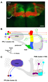
Figure 2. Zones of the mushroom body innervated by dopaminergic neurons
A. Projection view of a subset of dopaminergic neurons in the Protocerebral Posterior Lateral (PPL) 1 cluster. A brain from a transgenic fly expressing a red fluorescent protein in MB neurons and a green fluorescent protein in MB-MP neurons that innervate the MB-heel and peduncle region (shown as green in panel B) and also the neurons shown as magenta in panel B that seem to project around the MB. Image adapted, with permission, from Ref. [11]. Scale bar 20µm. B. The PPL1 cluster. The Butcher’s cut illustration summarizes data from Refs. [11,22,25]. The image shows one neuron projecting to the tip of the α lobe (red), one to the tip of the α´ tip (orange), one to the upper stalk of the vertical lobes (MB-V1 neuron, yellow), one to the lower stalk and junction region (MB-MV1 neuron, blue) and one to the heel and distal peduncle (MB-MP neuron, green). In addition, PPL1 includes a neuron that innervates the anterior superior and inferior medial protocerebrum (magenta), that runs along a similar path to the MB-MP neurons but does not appear to innervate the MB, and another neuron that innervates the region behind the α lobe and sends another branch into the central complex (grey cell body, projection not shown for clarity). All neurons shown have a projection to a similar zone on the contralateral MB (dotted lines with arrowhead). The cell body locations are not stereotyped. Although the number of neurons in each class has not been exhaustively determined, there appear to be at least two MB-MP neurons per PPL1 cluster [11]. There are also likely to be multiple neurons in some of the other classes because 4 of the 12 neurons in PPL1 remain to be accounted for. C. The PPL2ab and Protocerebral Anterior Medial (PAM) clusters. The illustration summarizes data described in Refs. [22,25]. At least two neurons from the PPL2ab cluster innervate the ipsilateral MB calyx (brown and blue). Tyrosine hydroxylase staining suggests that DA neurons project to discrete zones of the horizontal β, β´ and γ MB lobes (marked with black dotted lines for β and β´ and orange for γ) but the only individual PAM neurons described so far ramify on the tip of the β lobe (MB-M3 neurons, cyan) and a similar zone on the contralateral MB (dotted line with arrowhead) or further along the β lobe (MB-MVP1 neurons, purple).
Figure 3
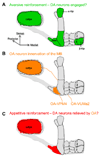
Figure 3. Hypothetical models for the involvement of dopaminergic (DA) neurons in aversive and appetitive reinforcement
A. DA neurons representing aversive reinforcement. Studies from Refs. [24,25] suggest that neurons in PPL1 convey negative reinforcement whereas live-imaging data from Ref. [22] indicates that DA neurons innervating the lower stalk, junction and α tip regions are most strongly activated by shock. In this illustration, a selection of DA neurons innervating these specific MB zones, and the MB-M3 neurons on the tip of the β lobe [25], are activated (green). B. Octopaminergic (OA) neuron innervation of the mushroom body (MB). Single cell analysis has determined that OA-Ventral Unpaired Medial (VUM)a2 neurons innervate the MB calyx whereas OA-Ventral Paired Medial (VPM) 4 neurons innervate the MB heel [23]. Should the OA neurons that innervate the MB be those that are important for appetitive reinforcement in Drosophila, they are appropriately placed to influence the activity of the DA neurons in the calyx and in the MB heel (the MB-MP neurons). C. DA neurons that could be relieved by appetitive reinforcement. OA neuron activation can substitute for sugar presentation in appetitive conditioning in Drosophila larvae [75] and in the honeybee [76]. In this model, OA neuron action would relieve the DA neurons in the heel (MB-MP neurons) and those in the calyx that can convey negative signals (now red rather than green in panel A). Hunger inhibits the MB-MP DA neurons to promote appetitive memory retrieval [11]. Therefore, learning could involve a similar MB-MP relief mechanism that relies on OA.

Similar articles

Cited by

References

    1. Joshua M, et al. The dynamics of dopamine in control of motor behavior. Curr Opin Neurobiol. 2009;19:615–620. - PubMed
    1. Dayan P, Balleine BW. Reward, motivation, and reinforcement learning. Neuron. 2002;36:285–298. - PubMed
    1. Wise RA. Dopamine, learning and motivation. Nat Rev Neurosci. 2004;5:483–494. - PubMed
    1. Montague PR, et al. Computational roles for dopamine in behavioural control. Nature. 2004;431:760–767. - PubMed
    1. Heinz A, Schlagenhauf F. Dopaminergic dysfunction in schizophrenia: salience attribution revisited. Schizophr Bull. 2010;36:472–485. - PMC - PubMed

Publication types

LinkOut - more resources