. 2010 Oct 22;285(43):33092-33103.
doi: 10.1074/jbc.M110.130377. Epub 2010 Aug 11.

CCAAT/enhancer-binding protein β and NF-κB mediate high level expression of chemokine genes CCL3 and CCL4 by human chondrocytes in response to IL-1β

Affiliations

CCAAT/enhancer-binding protein β and NF-κB mediate high level expression of chemokine genes CCL3 and CCL4 by human chondrocytes in response to IL-1β

Zhiqi Zhang et al. J Biol Chem. .

Abstract

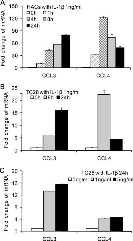
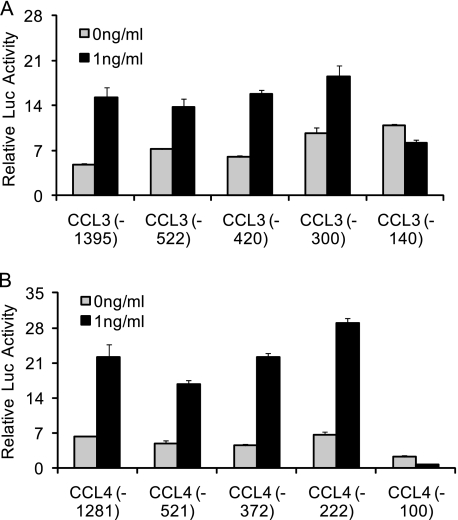
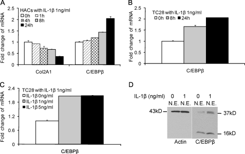
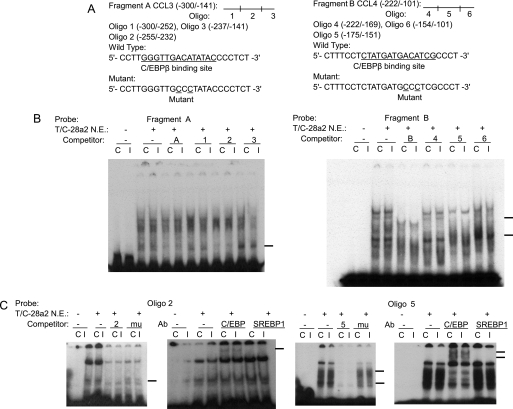
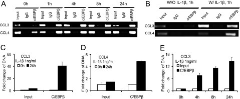
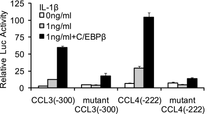
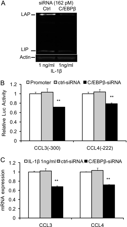
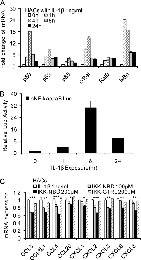
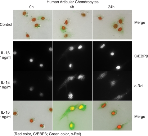
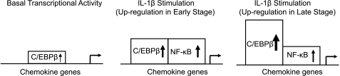
A large set of chemokines is highly up-regulated in human chondrocytes in response to IL-1β (Sandell, L. J., Xing, X., Franz, C., Davies, S., Chang, L. W., and Patra, D. (2008) Osteoarthr. Cartil. 16, 1560-1571). To investigate the mechanism of transcriptional regulation, deletion constructs of selected chemokine gene promoters, the human CCL3 (MIP-1α) and CCL4 (MIP-1β), were transfected into human chondrocytes with or without IL-1β. The results show that an IL-1β-responsive element is located between bp -300 and -140 of the CCL3 promoter and between bp -222 and -100 of the CCL4 promoter. Because both of these elements contain CCAAT/enhancer-binding protein β (C/EBPβ) motifs, the function of C/EBPβ was examined. IL-1β stimulated the expression of C/EBPβ, and the direct binding of C/EBPβ to the C/EBPβ motif was confirmed by EMSA and ChIP analyses. The -300 bp CCL3 promoter and -222 bp CCL4 promoter were strongly up-regulated by co-transfection with the C/EBPβ expression vector. Mutation of the C/EBPβ motif and reduction of C/EBPβ expression by siRNA decreased the up-regulation. Additionally, another cytokine-related transcription factor, NF-κB, was also shown to be involved in the up-regulation of chemokines in response to IL-1β, and the binding site was identified. The regulation of C/EBPβ and NF-κB was confirmed by the inhibition by C/EBPβ and NF-κB and by transfection with C/EBPβ and NF-κB expression vectors in the presence or absence of IL-1β. Taken together, our results suggest that C/EBPβ and NF-κB are both involved in the IL-1β-responsive up-regulation of chemokine genes in human chondrocytes. Time course experiments indicated that C/EBPβ gradually and steadily induces chemokine up-regulation, whereas NF-κB activity was highest at the early stage of chemokine up-regulation.


Figures

FIGURE 1.
FIGURE 1.
IL-1β stimulates the expression of CCL3 and CCL4 in normal human articular chondrocytes (HACs) and T/C-28 (TC28) chondrocyte cells. A, human articular chondrocytes were treated with 1 ng/ml IL-1β for various times as indicated. B, T/C-28 cells were treated with 1 ng/ml IL-1β for various times as indicated. C, T/C-28 cells were treated for various concentrations for 24 h. The relative expression levels were examined by the quantitative real time PCR method. Each bar represents the mean ± S.D. (error bars) from three experiments.
FIGURE 2.
FIGURE 2.
IL-1β-responsive element is located between −300 and −140 bp of the CCL3 promoter and between −222 and −100 bp of the CCL4 promoter. 5′-Deletion constructs were transiently transfected into T/C-28a2 cells and incubated for 24 h and then for a further 24 h (CCL3) (A) or 8 h (CCL4) (B) in the absence or presence of IL-1β (1 ng/ml) with fresh complete medium. Luciferase activities were measured and expressed relative to the activity of promoterless pGL3b (set as 1). Each bar represents the mean ± S.D. of at least three independent experiments.
FIGURE 3.
FIGURE 3.
IL-1β stimulates the expression of C/EBPβ in normal human articular chondrocytes (HACs) and T/C-28 chondrocyte cells. The relative expression levels were examined in normal chondrocytes from human articular cartilage and T/C-28 cells treated with IL-1β (1 ng/ml) using the quantitative real-time PCR method. A and B, cells were treated with 1 ng/ml IL-1β for various times as indicated in human articular chondrocytes (A) and T/C-28 cells (B). C, T/C-28 cells were treated with various concentrations of IL-1β for 24 h. D, C/EBPβ proteins were examined by Western blot of nuclear extracts (N.E.) from T/C-28 cells treated or without IL-1β (1 ng/ml). IL-1β increased all of the isoforms of C/EBPβ, LAP (38 and 36 kDa), and LIP (16 kDa), and both isoforms of C/EBPβ-LAP were more significantly increased than LIP. Each bar represents the mean ± S.D. from 3–5 experiments.
FIGURE 4.
FIGURE 4.
C/EBPβ binds the sequence between −251 and −238 bp of the CCL3 promoter and between −168 and −155 bp of the CCL4 promoter. A, a diagram showing DNA fragments used for EMSA. For CCL3, Fragment A from −300 to −141 bp and the relative locations of oligonucleotides 1, 2, and 3 are shown. For CCL4, Fragment B from −222 to −101 bp and the relative locations of oligonucleotides 4, 5, and 6 are shown. Oligonucleotide 2 from −255 to −232 bp and oligonucleotide 5 from −175 to −151 bp contain the C/EBPβ motif (underlined). Mu, mutant oligonucleotides 2 and 5 containing two base pair mutations (AA to CC) is underlined within the C/EBPβ motif. B, EMSA for T/C-28a2 nuclear extracts using Fragment A or B as a probe and various cold competitors at a 100-fold molar excess. Oligonucleotides 2 and 5 competed with the binding of nuclear proteins to the probe. C, EMSA and supershift analysis for T/C-28a2 nuclear extracts using oligonucleotides 2 and 5 as the probe. Mutant oligonucleotides 2 and 5 did not compete with the binding of nuclear proteins to the probe. Antibodies against C/EBPβ or SREBP1 were preincubated with the nuclear extracts. The lines show the shift band of the complex of C/EBPβ antibody (Ab) and specific retarded bands (C, control; I, IL-1β treatment).
FIGURE 5.
FIGURE 5.
C/EBPβ binding activity to the CCL3(−300/−141) and CCL4(−222/−101) promoter regions is increased in the presence of IL-1β. A chromatin immunoprecipitation assay was performed using anti-C/EBPβ antibodies in T/C-28 cells treated with IL-1β for various times as indicated. Treatment with IL-1β enhanced the binding of C/EBPβ to the promoter. A, T/C-28 cells were treated with 1 ng/ml IL-1β for 0, 1, 4, 8, and 24 h. B, T/C-28 cells were treated with or without IL-1β (1 ng/ml) for 1 h. C and D, T/C-28 cells were treated with 1 ng/ml IL-1β for 0 and 24 h, and the relative expression levels of input and C/EBPβ antibody DNAs of CCL3 (C) and CCL4 (D) were examined by the quantitative real time PCR method. The value was normalized to GAPDH, and the expression levels of input and C/EBPβ at 0 h were set as 1. E, T/C-28 cells were treated with 1 ng/ml IL-1β for 0, 4, 8, and 24 h, and the relative expression levels of CCL3 of input and C/EBPβ antibody DNAs were examined by the quantitative real-time PCR method. The value was normalized to GAPDH and input, and the expression levels of C/EBPβ at 0 h were set as 1. Error bars, S.D.
FIGURE 6.
FIGURE 6.
C/EBPβ functions as an activator for the CCL3 and CCL4 promoter activities, and mutation of the C/EBP-binding site down-regulates the promoter activity of the CCL3(−300/−1) and CCL4(−222/−1) constructs. Site-directed mutagenesis was performed within the C/EBP-binding site of the CCL3(−300/−1) and CCL4(−222/−1) constructs using the mutant oligonucleotide 2 and 5 sequence. The mutant and wild type constructs were transiently transfected into T/C-28a2 cells and incubated with or without IL-1β. Luciferase activities were measured and expressed relative to the activity of promoterless pGL3b (set as 1). Further, the wild type and mutant of CCL3(−300/−1) and CCL4(−222/−1) were co-transfected with C/EBPβ expression plasmid into T/C-28a2 cells with IL-1β (1 ng/ml). Relative luciferase activity indicates the -fold expression relative to the activity of promoterless pGL3b, which co-transfected with empty vector (set as 1) in the presence of IL-1β (1 ng/ml). Each bar represents the mean ± S.D. (error bars) of at least three independent experiments.
FIGURE 7.
FIGURE 7.
Silencing of C/EBPβ suppresses the IL-1β-induced CCL3 and CCL4 transcriptional activation. A, Odyssey Western blot of C/EBPβ proteins in T/C-28 cells transfected with control (Ctrl) or C/EBPβ siRNAs (162 pm), and stimulated with 1 ng/ml IL-1β for 8 h. B, T/C-28 cells were transfected with CCL3 and CCL4 promoters and, where indicated, cotransfected with control (ctrl) or C/EBPβ siRNAs (162 pm). Twenty-four h after transfection, T/C-28 cells were stimulated with 1 ng/ml IL-1β for 24 h (CCL3) or 8 h (CCL4). Relative luciferase activity indicates the -fold expression relative to the activity of promoter with IL-1β (1 ng/ml) only (set as 1). C, T/C-28 cells transfected with control or C/EBPβ siRNAs were stimulated for 8 h with IL-1β (1 ng/ml). The relative expression levels were examined by the quantitative real time PCR method. The p value of C/EBPβ siRNA was compared with IL-1β alone based on Student's t test (**, p < 0.01). Each bar represents the mean ± S.D. (error bars) of at least three independent experiments.
FIGURE 8.
FIGURE 8.
Involvement of NF-κB in the up-regulation of chemokines in human chondrocytes in response to IL-1β. A, human articular chondrocytes were treated with 1 ng/ml IL-1β for various times as indicated. B, effect of IL-1β (1 ng/ml) on the expression of pNF-κB Luc reporter in T/C-28 human chondrocytes. Relative luciferase activity indicates the -fold expression relative to the activity of zero time (set as 1) in the presence of IL-1β (1 ng/ml). C, human articular chondrocytes were pretreated with vehicle (DMSO), IKK-NBD peptide (100 and 200 μm), or IKK-NBD control peptide (200 μm) for 1 h and then exposed to IL-1β (1 ng/ml) for 4 h. The p value of IKK-NBD (200 μm) was compared with IL-1β alone based on Student's t test (*, p < 0.05; **, p < 0.01; ***, p < 0.001). Each bar represents the mean ± S.D. (error bars) of three independent experiments.
FIGURE 9.
FIGURE 9.
Transcriptional regulation in the highly expression of CCL3 and CCL4. A, the candidate C/EBPβ and NF-κB binding sites in human CCL3 (−1395) and CCL4 (−1281) constructs. B, C/EBPβ and IKK2 stimulate the expression of CCL3 (−1395) and CCL4 (−1281) promoter in T/C-28 human chondrocytes. The CCL3 (−1395) and CCL4 (−1281) promoter constructs were co-transfected with C/EBPβ and IKK2 expression plasmid into T/C-28a2 cells with or without IL-1β (1 ng/ml) for 24 h (CCL3) or 8 h (CCL4). Relative luciferase activity indicates the -fold expression relative to the activity of the construct co-transfected with empty vector (set as 1) in the presence of IL-1β after each co-transfection compared with their activities in the absence of IL-1β. Each bar represents the mean ± S.D. (error bars) of three independent experiments.
FIGURE 10.
FIGURE 10.
Involvement of NF-κB in IL-1β-responsive elements of chemokines in human chondrocytes. The CCL3 (−300) (A), CCL3 (−140) (A), CCL4 (−222) (B), and CCL4 (−100) (B) promoter constructs were co-transfected into T/C-28a2 cells. Twenty-four h after transfection, T/C-28 cells were pretreated with vehicle (DMSO), IKK-NBD peptide (100 μm), or IKK-NBD control peptide (100 μm) for 1 h and then exposed to IL-1β (1 ng/ml) for 4 h. The p value of IKK-NBD (100 μm) was compared with IL-1β alone based on Student's t test (**, p < 0.01; ***, p < 0.001). Each bar represents the mean ± S.D. (error bars) of three independent experiments.
FIGURE 11.
FIGURE 11.
IKK-NBD and SB303580 (SB) co-enhance the inhibition of chemokines by human chondrocytes with IL-1β treatment. A, T/C-28 cells were pretreated with vehicle (DMSO), IKK-NBD peptide (100 μm), SB303580 (100 μm), or IKK-NBD control peptide (100 μm) for 1 h and then exposed to IL-1β (1 ng/ml) for 4 h. After IL-1β treatment, total RNA was isolated, and real-time quantitative PCR was performed. B, the CCL3 (−1395) and CCL4 (−1281) promoter constructs were transfected into T/C-28a2 cells and incubated for 24 h and then were pretreated with vehicle (DMSO), IKK-NBD peptide (100 μm), SB303580 (100 μm), and IKK-NBD control peptide (100 μm) for 1 h and exposed to IL-1β (1 ng/ml) for 4 h. Relative luciferase activity indicates the -fold expression relative to the activity of the construct co-transfected with empty vector (set as 1) in the presence of IL-1β (1 ng/ml). The p value of IKK-NBD (100 μm) or SB303580 (100 μm) was compared with IL-1β alone based on Student's t test (**, p < 0.01; ***, p < 0.001). Each bar represents the mean ± S.D. (error bars) of at least three independent experiments.
FIGURE 12.
FIGURE 12.
Subcellular localization of C/EBPβ and c-Rel in response to IL-1β. The top panel shows a merged image of immunohistochemistry for C/EBPβ and the NF-κB subunit c-Rel; C/EBPβ is present in the nucleus of cells even without IL-1β exposure, whereas c-Rel is located diffusely throughout the cell. With the addition of IL-1β, C/EBPβ is increased in the nucleus, and c-Rel is increased and translocates to the nucleus. At 24 h, C/EBPβ remains high in the nucleus, and c-Rel is significantly reduced.
FIGURE 13.
FIGURE 13.
A transcriptional model for the up-regulation of chemokine genes in human chondrocytes in response to IL-1β. Upon IL-1β stimulation, C/EBPβ protein and DNA binding are increased up to the 24 h tested. NF-κB showed little function without IL-1β but increased up to 8 h, after which the function was greatly reduced. The distribution of the transcription factors (see Fig. 12) is consistent with the functional parameters.

Similar articles

Cited by

References

    1. Gerard C., Rollins B. J. (2001) Nat. Immunol. 2, 108–115 - PubMed
    1. Haringman J. J., Ludikhuize J., Tak P. P. (2004) Ann. Rheum. Dis. 63, 1186–1194 - PMC - PubMed
    1. Ma Q., Jones D., Borghesani P. R., Segal R. A., Nagasawa T., Kishimoto T., Bronson R. T., Springer T. A. (1998) Proc. Natl. Acad. Sci. U.S.A. 95, 9448–9453 - PMC - PubMed
    1. Acosta J. C., O'Loghlen A., Banito A., Guijarro M. V., Augert A., Raguz S., Fumagalli M., Da Costa M., Brown C., Popov N., Takatsu Y., Melamed J., d'Adda di Fagagna F., Bernard D., Hernando E., Gil J. (2008) Cell 133, 1006–1018 - PubMed
    1. Han J. H., Choi S. J., Kurihara N., Koide M., Oba Y., Roodman G. D. (2001) Blood 97, 3349–3353 - PubMed

Publication types

MeSH terms

LinkOut - more resources