. 2010 Oct 15;285(42):32504-11.
doi: 10.1074/jbc.M110.153080. Epub 2010 Aug 10.

Structure and metal loading of a soluble periplasm cuproprotein

Affiliations

Structure and metal loading of a soluble periplasm cuproprotein

Kevin J Waldron et al. J Biol Chem. .

Abstract

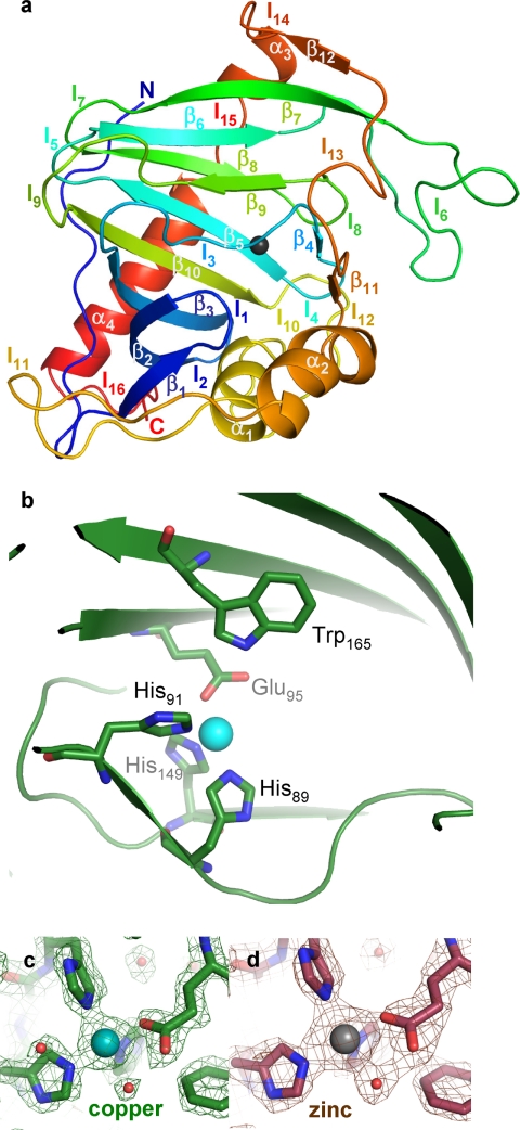
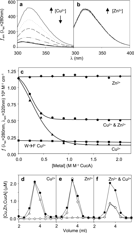
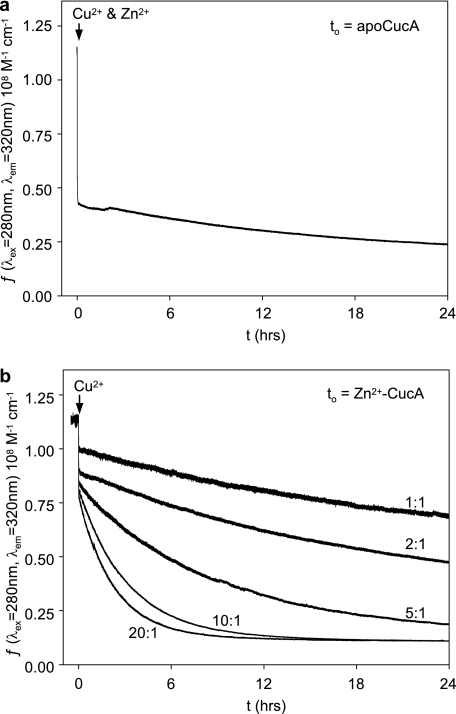
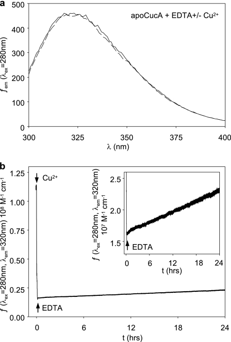
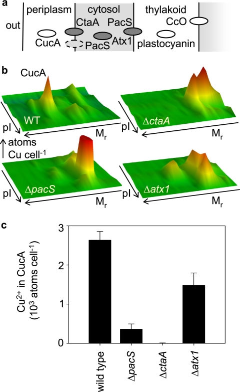
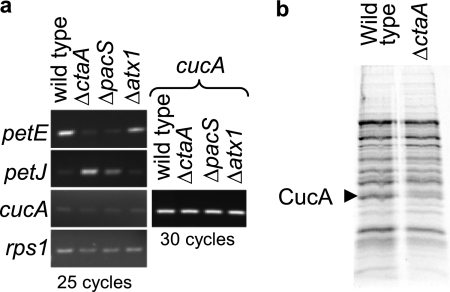
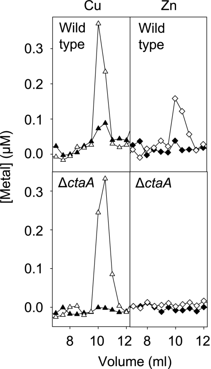
A copper-trafficking pathway was found to enable Cu(2+) occupancy of a soluble periplasm protein, CucA, even when competing Zn(2+) is abundant in the periplasm. Here, we solved the structure of CucA (a new cupin) and found that binding of Cu(2+), but not Zn(2+), quenches the fluorescence of Trp(165), which is adjacent to the metal site. Using this fluorescence probe, we established that CucA becomes partly occupied by Zn(2+) following exposure to equimolar Zn(2+) and Cu(2+). Cu(2+)-CucA is more thermodynamically stable than Zn(2+)-CucA but k((Zn→Cu)exchange) is slow, raising questions about how the periplasm contains solely the Cu(2+) form. We discovered that a copper-trafficking pathway involving two copper transporters (CtaA and PacS) and a metallochaperone (Atx1) is obligatory for Cu(2+)-CucA to accumulate in the periplasm. There was negligible CucA protein in the periplasm of ΔctaA cells, but the abundance of cucA transcripts was unaltered. Crucially, ΔctaA cells overaccumulate low M(r) copper complexes in the periplasm, and purified apoCucA can readily acquire Cu(2+) from ΔctaA periplasm extracts, but in vivo apoCucA fails to come into contact with these periplasmic copper pools. Instead, copper traffics via a cytoplasmic pathway that is coupled to CucA translocation to the periplasm.


Figures

FIGURE 1.
FIGURE 1.
Crystal structure of Cu2+-CucA. a, structural elements (α, α-helices; β, β-strands; l, loops), colored in order from the amino (blue) to carboxyl (red) terminus, are numerically labeled. b, residues (green) surrounding the copper atom (blue sphere) of Cu2+-CucA are shown in the closed conformation. c, residues surrounding the metal atoms within CucA with corresponding composite omit electron density maps each contoured at 1.5 σ for Cu2+-CucA, and d, Zn2+-CucA.
FIGURE 2.
FIGURE 2.
Copper and zinc both bind to CucA. a, quenching of the fluorescence emission spectra of CucA (5 μm in 10 mm HEPES, pH 7.0, 150 mm NaCl) exposed to increasing concentrations of Cu2+, 280 nm excitation. b, lack of quenching of the fluorescence emission spectra of CucA, as in a, exposed to increasing concentrations of Zn2+. c, fluorescence emission of CucA at 320 nm as a function of added Zn2+ (diamonds), Cu2+ (triangles), and simultaneous addition of equimolar Zn2+ plus Cu2+ to 5 μm apoCucA (circles). Fluorescence was measured 60 s after metal addition and mixing. Fluorescence emission of a CucA W165F variant was also determined (closed squares). Fractionation of CucA-bound metal by size exclusion chromatography with metal (open diamonds, zinc; open triangles, copper) detected by ICP-MS after addition of 10 μm Cu2+ (d), 10 μm Zn2+ (e), and 10 μm Zn2+ plus 10 μm Cu2+ (f) to 5 μm apoCucA (closed circles, protein).
FIGURE 3.
FIGURE 3.
Zn2+-CucA exchanges slowly with Cu2+. a, quenching of the fluorescence emission of apoCucA (5 μm) as a function of time following simultaneous addition of equimolar Zn2+ and Cu2+ (both at 2.5 m excess with respect to CucA). b, quenching of the fluorescence emission of Zn2+-CucA (2.5 m excess relative to CucA) as a function of time following addition of Cu2+ at increasing molar eq relative to Zn2+ (1:1, 2:1, 5:1, 10:1, and 20:1).
FIGURE 4.
FIGURE 4.
Dissociation of Cu2+-CucA is slow. a, intrinsic fluorescence of CucA (5 μm) is not quenched by 2.5 m excess Cu2+ added in the presence of EDTA (1 mm). b, slow restoration of intrinsic fluorescence of CucA after initial quenching with 2.5 m eq of Cu2+ followed by the addition of excess EDTA (1 mm). Inset is plotted on an expanded y axis to show the rate of Cu2+-CucA dissociation.
FIGURE 5.
FIGURE 5.
CucA obtains less Cu2+ in trafficking mutants. a, cuproproteins CucA, plastocyanin and cytochrome oxidase (CcO), are located in the periplasm and thylakoid compartments, respectively. Copper metallochaperone Atx1 is cytosolic and interacts with amino-terminal regions of copper ATPases CtaA and PacS. PacS is predominantly located in thylakoid membranes. b, profile of periplasmic copper complexes from wild-type and mutant cells, separated using two-dimensional liquid chromatography based on relative molecular mass (Mr) and charge (pI). c, number of atoms of Cu2+ cell−1 bound to CucA in wild-type and mutant cells calculated from the volumes under CucA peaks (as in b). Data are the means of three independent replicates with S.D. of triplicates.
FIGURE 6.
FIGURE 6.
Loss of CucA protein but not transcripts in Cu2+-trafficking mutants. a, RT-PCRs using RNA isolated from the four genotypes and primers corresponding to the four transcripts as shown. All products are reverse transcriptase-dependent (supplemental Fig. 5). 25 cycles, plus 30 cycles for cucA, were used. Similar trends were observed in two further experiments using independent RNA isolates. b, SDS-PAGE of crude periplasm extracts of wild-type and ΔctaA mutants after concentration using ion exchange chromatography.
FIGURE 7.
FIGURE 7.
In vitro apoCucA acquires copper from ΔctaA but zinc and copper from wild-type periplasm extracts. Purified CucA protein (1 ml of 1.5 μm in 10 mm HEPES, pH 7.0, 150 mm NaCl) (open symbols) or buffer control (closed symbols) was added to periplasm extracts, which were then concentrated by anion exchange and eluted metal complexes resolved by gel filtration HPLC (TSK SW-3000) and eluant analyzed for copper (triangles) and zinc (diamonds) by ICP-MS.

References

    1. Nies D. H. (2007) in Molecular Microbiology of Heavy Metals (Nies D. H., Silver S. eds) pp. 117–142, Springer-Verlag, Heidelberg, Germany
    1. Nikaido H., Vaara M. (1985) Microbiol. Rev. 49, 1–32 - PMC - PubMed
    1. Tottey S., Waldron K. J., Firbank S. J., Reale B., Bessant C., Sato K., Cheek T. R., Gray J., Banfield M. J., Dennison C., Robinson N. J. (2008) Nature 455, 1138–1142 - PubMed
    1. Santini C. L., Ize B., Chanal A., Müller M., Giordano G., Wu L. F. (1998) EMBO J. 17, 101–112 - PMC - PubMed
    1. Palmer T., Berks B. C. (2003) Microbiology 149, 547–556 - PubMed

Publication types

MeSH terms

LinkOut - more resources