. 2010 Sep 15;16(18):4583-94.
doi: 10.1158/1078-0432.CCR-10-0733. Epub 2010 Aug 11.

A novel chemoimmunomodulating property of docetaxel: suppression of myeloid-derived suppressor cells in tumor bearers

Affiliations

A novel chemoimmunomodulating property of docetaxel: suppression of myeloid-derived suppressor cells in tumor bearers

Krithika N Kodumudi et al. Clin Cancer Res. .

Abstract

Purpose: Myeloid-derived suppressor cells (MDSC) accumulate in tumor-bearing hosts and are associated with immune suppression. To date, there have only been few studies that evaluate the direct effect of chemotherapeutic agents on MDSCs. Agents that inhibit MDSCs may be useful in the treatment of patients with various cancers.

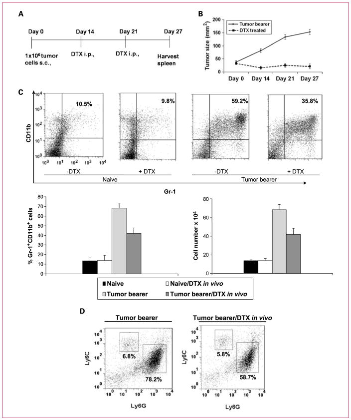
Experimental design: We investigated the in vivo effects of docetaxel on immune function in 4T1-Neu mammary tumor-bearing mice to examine if a favorable immunomodulatory effect accompanies tumor suppression. Primary focus was on the differentiation status of MDSCs and their ability to modulate T-cell responses.

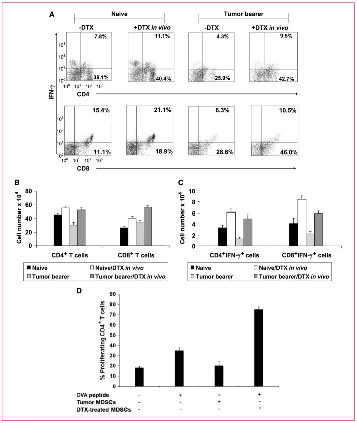
Results: Docetaxel administration significantly inhibited tumor growth in 4T1-Neu tumor-bearing mice and considerably decreased MDSC proportion in the spleen. The treatment also selectively increased CTL responses. Docetaxel-pretreated MDSCs cocultured with OT-II splenocytes in the presence of OVA(323-339) showed OT-II-specific CD4 activation and expansion in vitro. In characterizing the phenotype of MDSCs for M1 (CCR7) and M2 [mannose receptor (CD206)] markers, MDSCs from untreated tumor bearers were primarily MR(+) with few CCR7(+) cells. Docetaxel treatment polarized MDSCs toward an M1-like phenotype, resulting in 40% of MDSCs expressing CCR7 in vivo and in vitro, and macrophage differentiation markers such as MHC class II, CD11c, and CD86 were upregulated. Interestingly, docetaxel induced cell death selectively in MR(+) MDSCs while sparing the M1-like phenotype. Finally, inhibition of signal transducer and activator of transcription 3 may in part be responsible for the observed results.

Conclusions: These findings suggest potential clinical benefit for the addition of docetaxel to current immunotherapeutic protocols.


Conflict of interest statement

Disclosure of Potential Conflicts of Interest

No potential conflicts of interest were disclosed.

Figures

Fig. 1
Fig. 1
Docetaxel treatment of BALB/c mice bearing established 4T1 mammary tumors reduces tumor growth. A, schematic representation of the docetaxel (DTX) treatment regimen. B, in vivo injection of docetaxel in tumor bearers retarded tumor growth (n = 10). C, docetaxel treatment (33 mg/kg body weight) reduced Gr-1+CD11b+ MDSCs in the spleens of mice bearing tumors. Spleen cells were stained with PE-conjugated anti–Gr-1 and APC-conjugated anti-CD11b antibodies. The percentages of double-positive Gr-1+CD11b+ cells are shown for spleens of three mice per group as follows: (a) naive mice, (b) docetaxel-treated naive mice, (c) untreated tumor bearers, and (d) docetaxel-treated tumor bearers. Columns, mean of three separate experiments; bars, SE. D, docetaxel treatment reduced granulocytic MDSCs. Monocytic MDSCs are Ly6ChiLy6Glo, whereas granulocytic MDSCs are Ly6CloLy6Ghi.
Fig. 2
Fig. 2
Docetaxel treatment of tumor-bearing mice upregulates CD4+ and CD8+ T cells and generates significant IFN-γ production. A, flow cytometric analysis of cells isolated from spleens of naive mice, docetaxel-treated naive mice, tumor bearers, and docetaxel-treated tumor bearers. Dual staining for surface CD4 or CD8 and intracellular IFN-γ showed that both CD4 and CD8 T cells had increased intracellular IFN-γ after docetaxel treatment. B, columns, mean cell number of CD4+ and CD8+ T cells (n = 3); bars, SE. C, columns, mean cell number of IFN-γ–producing cells (n = 3); bars, SE. D, docetaxel treatment causes MDSCs to lose their suppressive effect on CD4+ T cells and significantly increased T-cell–specific responses to antigen. Splenocytes from OT-II transgenic mice were stimulated with OVA323–329 peptide in the presence of docetaxel-treated or untreated MDSCs. In vitro proliferation of CD4+ T cells was measured by CFSE staining after 24 h. Columns, mean percentage of CD4+ T-cell–proliferating cells; bars, SE.
Fig. 3
Fig. 3
Spleen cells from docetaxel-treated tumor-bearing mice have cytotoxic effect on 4T1 tumor cells. A 24-h 51Cr release assay was done using 4T1 tumor cells as targets and T cells from tumor bearers, either treated or untreated with docetaxel in vivo. The T cells were purified using a commercial affinity column and were >95% pure. The percentage of specific 51Cr release was determined by the following equation: (experimentalcpm-spontaneouscpm)/(totalcpmincorporated-spontaneouscpm)×100. All determinations were done in triplicate, and the SEM of all assays was calculated and was typically 5% of the mean or less.
Fig. 4
Fig. 4
A, MDSCs from docetaxel-treated mice express M1 markers. MDSCs were affinity column purified from three mice per group representing naive mice, docetaxel-treated naive mice, tumor bearers, and docetaxel-treated tumor bearers. They were immunostained for CCR7 and iNOS to represent M1 markers or MR to represent a specific M2 marker before flow cytometric analysis. A, histogram representation of MR, CCR7, and iNOS levels in naive mice, docetaxel-treated naive mice, tumor bearers, and docetaxel-treated tumor bearers. B, columns, mean fluorescence intensity units of CCR7, MR, and iNOS from three independent experiments; bars, SE. C, in vivo docetaxel-treated MDSCs have increased differentiation markers. MACS column–purified MDSCs from the above treatment groups were analyzed by flow cytometry for MHC class II, CD11c, CD40, CD80, and CD85 expression. Gray filled histograms are isotype controls, dotted lines represent the docetaxel-treated MDSCs, and black solid lines represent untreated MDSCs.
Fig. 5
Fig. 5
Docetaxel differentiates MDSC into M1-like phenotype in vitro and selectively inhibits M2-like phenotype of MDSCs. A, MACS column–purified MDSCs were assayed by flow cytometry, confirmed to be >90% Gr-1+CD11b+ cells, and further used for in vitro assays. MDSCs were cultured in the presence or absence of docetaxel at 11 nmol/L for 24 h followed by analysis for M1 (CCR7) and M2 (MR) markers. A, flow cytometric analysis of the percentage of CCR7- and MR-expressing MDSCs before and after docetaxel culture. B, flow cytometric analysis of Annexin V+ MDSCs before and after docetaxel culture. C and D, percentage of Annexin V+ and Annexin V cells in CCR7+ or MR+ MDSCs. Docetaxel induced apoptosis on MDSCs, and the cells that were differentiated into M1-like phenotype were negative for Annexin V but positive for CCR7. E, cytokine analysis of supernatants of MDSCs cultured in the presence or absence of docetaxel for 24 h. Docetaxel-treated MDSCs enhance IL-12 and suppress IL-10 production.
Fig. 6
Fig. 6
Docetaxel selectively inhibits MDSCs via STAT3 signaling. A and B, lysates from MACS column–purified MDSCs were immunoblotted (IB) for both phosphorylated and total AKT, STAT1, and STAT3. MDSCs were treated or untreated with docetaxel (11 nmol/L) or JSI 124 (0.125 μmol/L) for 0, 3, and 6 h before analysis of phosphoproteins. C and D, MDSCs, treated or untreated with JSI 124, were immunostained for CCR7 and MR markers followed by Annexin V staining. Columns, mean fluorescence intensity of the Annexin V+ and Annexin V CCR7- and MR-expressing cells. E, cytokine analysis of supernatants of MDSCs cultured in the presence or absence of JSI 124 for 24 h. JSI 124–treated MDSCs enhanced IL-12 and suppressed IL-10 production.

References

    1. Gabrilovich DI, Nagaraj S. Myeloid-derived suppressor cells as regulators of the immune system. Nat Rev Immunol. 2009;9:162–74. - PMC - PubMed
    1. Kusmartsev S, Gabrilovich DI. Role of immature myeloid cells in mechanisms of immune evasion in cancer. Cancer Immunol Immunother. 2006;55:237–45. - PMC - PubMed
    1. Serafini P, De Santo C, Marigo I, et al. Derangement of immune responses by myeloid suppressor cells. Cancer Immunol Immunother. 2004;53:64–72. - PMC - PubMed
    1. Terabe M, Matsui S, Park JM, et al. Transforming growth factor-β production and myeloid cells are an effector mechanism through which CD1d-restricted T cells block cytotoxic T lymphocyte-mediated tumor immunosurveillance: abrogation prevents tumor recurrence. J Exp Med. 2003;198:1741–52. - PMC - PubMed
    1. Youn JI, Nagaraj S, Collazo M, Gabrilovich DI. Subsets of myeloid-derived suppressor cells in tumor-bearing mice. J Immunol. 2008;181:5791–802. - PMC - PubMed

Publication types

MeSH terms