. 2010 Oct;84(20):10467-76.
doi: 10.1128/JVI.00983-10. Epub 2010 Aug 11.

Viral cell death inhibitor MC159 enhances innate immunity against vaccinia virus infection

Affiliations

Viral cell death inhibitor MC159 enhances innate immunity against vaccinia virus infection

Sreerupa Challa et al. J Virol. 2010 Oct.

Abstract

Viral inhibitors of host programmed cell death (PCD) are widely believed to promote viral replication by preventing or delaying host cell death. Viral FLIPs (Fas-linked ICE-like protease [FLICE; caspase-8]-like inhibitor proteins) are potent inhibitors of death receptor-induced apoptosis and programmed necrosis. Surprisingly, transgenic expression of the viral FLIP MC159 from molluscum contagiosum virus (MCV) in mice enhanced rather than inhibited the innate immune control of vaccinia virus (VV) replication. This effect of MC159 was specifically manifested in peripheral tissues such as the visceral fat pad, but not in the spleen. VV-infected MC159 transgenic mice mounted an enhanced innate inflammatory reaction characterized by increased expression of the chemokine CCL-2/MCP-1 and infiltration of γδ T cells into peripheral tissues. Radiation chimeras revealed that MC159 expression in the parenchyma, but not in the hematopoietic compartment, is responsible for the enhanced innate inflammatory responses. The increased inflammation in peripheral tissues was not due to resistance of lymphocytes to cell death. Rather, we found that MC159 facilitated Toll-like receptor 4 (TLR4)- and tumor necrosis factor (TNF)-induced NF-κB activation. The increased NF-κB responses were mediated in part through increased binding of RIP1 to TNFRSF1A-associated via death domain (TRADD), two crucial signal adaptors for NF-κB activation. These results show that MC159 is a dual-function immune modulator that regulates host cell death as well as NF-κB responses by innate immune signaling receptors.


Figures
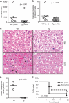
FIG. 1.
FIG. 1.
MC159 protects against LPS-induced liver injury. (A) Serum ALT level or (B) caspase-3 activity was determined 5 h after LPS plus d-galactosamine treatment. (C) Hematoxylin and eosin staining of liver sections of wild-type and MC159 transgenic mice treated with phosphate-buffered saline (PBS) or LPS for 5 h. Note that the extensive apoptotic nuclei in the wild-type hepatocytes (indicated by the arrows) were absent in the LPS-treated transgenic liver. Pictures shown are representative of images taken from two experiments with three different mice each. 200× magnification. (D) Decreased apoptosis in LPS-treated transgenic liver. The number of condensed apoptotic nuclei was counted in a double-blind manner. Each circle represents the average number of apoptotic nuclei from six different fields from one mouse. The results shown are representative of those of two experiments with at least three mice in each group. (E) Kaplan-Meier survival curve of LPS-treated wild-type and transgenic mice. WT, wild type; Tg, transgenic.
FIG. 2.
FIG. 2.
Enhanced clearance of VV in the peripheral tissues of MC159 transgenic mice. Wild-type and MC159 transgenic mice were intraperitoneally infected with 1 × 10⁶ PFU of WR VV. Viral titers per the whole tissue or organ were determined 4 days postinfection for the visceral fat pad (A), liver (B), and spleen (C) using a Vero cell plaque assay as described previously (8). The titers shown for different tissues were determined from the same group of mice. (D) Expression of the VV antigen E3L in infected tissues. Fat pads (lanes 1 to 6), livers (lanes 7 to 10), and spleens (lanes 11 to 14) were harvested from wild-type or transgenic mice 24 h postinfection. Tissue lysates were analyzed by Western blotting for expression of E3L, green fluorescent protein (GFP; for MC159-GFP), and β-actin as indicated. Uninfected tissues were included as controls as indicated. (E) Viral titers for wild-type transgenic mice infected with VV were determined 5 days after infection as in panels A to C.
FIG. 3.
FIG. 3.
Enhanced γδ T-cell infiltration in VV-infected MC159 transgenic mice. Hematoxylin and eosin staining of livers (A) and visceral fat pads (B) of uninfected wild-type control mice (a), VV-infected wild-type mice (b and d), or VV-infected transgenic mice (c and e). Tissues were harvested 24 h after infection. Panels a to c, 40× magnification; panels d and e, 200× magnification of the boxed area in panels b and c. The black arrows indicate the inflammatory cells. Images taken were representative of results of two experiments, each with four animals in each group. (C) Elevated expression of the chemokine CCL2 in the transgenic fat pad. The induction of CCL2 in VV-infected wild-type and transgenic fat pads was determined by quantitative PCR using 18S RNA as the internal control. Fold induction was determined by comparing the expression in infected mice with that in uninfected mice. (D) Transgenic and wild-type mice exhibited similar induction of IL-6 upon VV infection. IL-6 expression in visceral fat pads was determined as in panel C. (E and F) Enhanced γδ T-cell infiltration in the visceral fat pads of MC159 transgenic mice upon VV infection. Fat pad lymphocytes were isolated from wild-type (a) or transgenic (b) littermates 24 h after VV infection. (E) VV-infected transgenic fat pad lymphocytes were enriched in CD3+ T cells. Fat pad lymphocytes were harvested by enzyme digestion and stained with CD3 and Ly6G/C. The numbers represent the percentages of cells in the respective quadrants. (F) Increased γδ T cells in VV-infected transgenic fat pad lymphocytes. CD3+ cells were gated and analyzed for the expression of γδ TCR. The numbers represent the percentages of γδ TCR+ T cells. (G and H) Increased cell death in the livers of VV-infected MC159 transgenic mice. (G) The serum ALT level was determined 3 days postinfection. (H) Increased caspase-3 activation in VV-infected MC159 transgenic mice. Liver cell extracts were harvested 24 h postinfection and analyzed for active caspase-3 using a fluorogenic substrate assay as described in Materials and Methods. (I and J) Fat pad lymphocytes from MC159 transgenic mice exhibited normal cell death markers during VV infection. (I) Fat pad lymphocytes were harvested 24 h after VV infection, incubated for 1 h at 37°C, and stained with CD3, γδ TCR, and TUNEL. The percentages of TUNEL-positive CD3+ γδ TCR+ cells are indicated. (J) Fat pad lymphocytes were harvested as for panel I and incubated at 37°C for 5 h prior to staining with CD3 and annexin V. The percentages of cells that were annexin V positive are indicated. FACS plots and histology were representative of those of at three experiments with three animals in each experiment.
FIG. 4.
FIG. 4.
MC159 promotes NF-κB activation in parenchymal and hematopoietic cells. (A) Wild-type and transgenic MEFs were stimulated with recombinant mouse TNF for the indicated times. IκBα phosphorylation and degradation were analyzed by Western blotting. β-Actin was used as the internal control. (B) MEFs were stimulated with 100 pg/ml of LPS. IκBα phosphorylation and degradation were analyzed as in panel A. (C) BMDCs were stimulated with 100 ng/ml LPS for the indicated times. IκBα degradation was monitored by Western blotting. β-Actin is shown as the control. (D) BMMs were stimulated with LPS for the indicated times. An EMSA was performed with nuclear extracts isolated from wild-type or transgenic BMMs. Results are representative of those of two experiments. NS, nonspecific band.
FIG. 5.
FIG. 5.
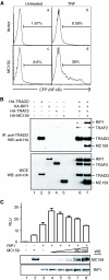
MC159 promotes NF-κB induction in a dose-dependent manner by facilitating the binding between RIP1 and TRADD. (A) MC159 enhances NF-κB activation in Jurkat cells. Jurkat cells were transfected with an NF-κB responsive CFP reporter and either an empty GFP vector (a and b) or MC159-GFP (c and d). Sixteen hours later, cells were stimulated with 10 ng/ml human recombinant TNF for 8 h. CFP expression as an indicator of NF-κB activity was analyzed in the transfected GFP+ population by flow cytometry. The numbers represent the percentages of CFP+ cells in the GFP+ populations. (B) MC159 enhances RIP1 binding to the TNFR-1 adaptor TRADD. HEK 293T cells were transfected with the indicated HA-tagged plasmids. TRADD immune complexes were isolated (top panel) with TRADD-specific antibody (immunoprecipitation [IP]). The presence of RIP1, TRAF2, and MC159 was examined by Western blotting (WB) with anti-HA antibody. The bottom panel shows the expression of the proteins in whole-cell extracts (WCE). (C) MC159 enhances RIP1-dependent NF-κB activation in a dose-dependent manner. HEK 293T cells were transfected with NF-κB-driven luciferase reporter, a constitutive active β-galactosidase reporter, a RIP1 expression plasmid, and increasing amounts of MC159 expression plasmid (lanes 4 to 8, 50 ng, 100 ng, 200 ng, 500 ng, and 800 ng, respectively). Luciferase activity was normalized to β-galactosidase activity and is shown as relative light units (RLU). The averages and standard errors of the means (SEM) from experiments performed in triplicate are shown. The bottom panels show the Western blot results for expression of RIP1 and MC159 in the corresponding samples. Results are representative of those of three experiments.
FIG. 6.
FIG. 6.
Expression of MC159 in the parenchyma, but not in the hematopoietic compartment, contributes to the enhanced γδ T-cell infiltration and control of viral replication. (A) Flow cytometric analysis of splenocytes isolated from radiation chimeras. Representative results from a Tg→wild-type chimera shows that >84% of lymphocytes were of the donor origin after reconstitution. (B) Radiation chimeras were generated as described in Materials and Methods. Four months after reconstitution with the indicated bone marrow cells, the mice were challenged with VV. Viral titers for the whole visceral fat pads were determined 3.5 days postinfection. (C) Radiation chimeras were created by transferring transgenic or wild-type bone marrow into wild-type recipient mice. Four months after bone marrow reconstitution, the chimeras and age-matched transgenic mice were challenged with VV. Fat pad lymphocytes were harvested 24 h postinfection and analyzed for the percentage of γδ TCR+ cells within the CD3+ population by flow cytometry. (D) Transgenic mice reconstituted with wild-type bone marrow cells and age-matched transgenic mice were analyzed for γδ T-cell infiltration to the visceral fat pads by flow cytometry 24 h post-VV infection. Results are representative of those of two experiments.

References

    1. Bahl, K., A. Huebner, R. J. Davis, and R. M. Welsh. 2010. Analysis of apoptosis of memory T cells and dendritic cells during the early stages of viral infection or exposure to toll-like receptor agonists. J. Virol. 84:4866-4877. - PMC - PubMed
    1. Balachandran, S., T. Venkataraman, P. B. Fisher, and G. N. Barber. 2007. Fas-associated death domain-containing protein-mediated antiviral innate immune signaling involves the regulation of Irf7. J. Immunol. 178:2429-2439. - PubMed
    1. Benedict, C. A., P. S. Norris, and C. F. Ware. 2002. To kill or be killed: viral evasion of apoptosis. Nat. Immunol. 3:1013-1018. - PubMed
    1. Bertin, J., R. C. Armstrong, S. Ottilie, D. A. Martin, Y. Wang, S. Banks, G. H. Wang, T. G. Senkevich, E. S. Alnemri, B. Moss, M. J. Lenardo, K. J. Tomaselli, and J. I. Cohen. 1997. Death effector domain-containing herpesvirus and poxvirus proteins inhibit both Fas- and TNFR1-induced apoptosis. Proc. Natl. Acad. Sci. U. S. A. 94:1172-1176. - PMC - PubMed
    1. Bukowski, J. F., B. A. Woda, S. Habu, K. Okumura, and R. M. Welsh. 1983. Natural killer cell depletion enhances virus synthesis and virus-induced hepatitis in vivo. J. Immunol. 131:1531-1538. - PubMed

Publication types

MeSH terms