. 2010 Oct;84(20):10644-52.
doi: 10.1128/JVI.00703-10. Epub 2010 Aug 11.

MicroRNA 203 expression in keratinocytes is dependent on regulation of p53 levels by E6

Affiliations

MicroRNA 203 expression in keratinocytes is dependent on regulation of p53 levels by E6

Declan J McKenna et al. J Virol. 2010 Oct.

Abstract

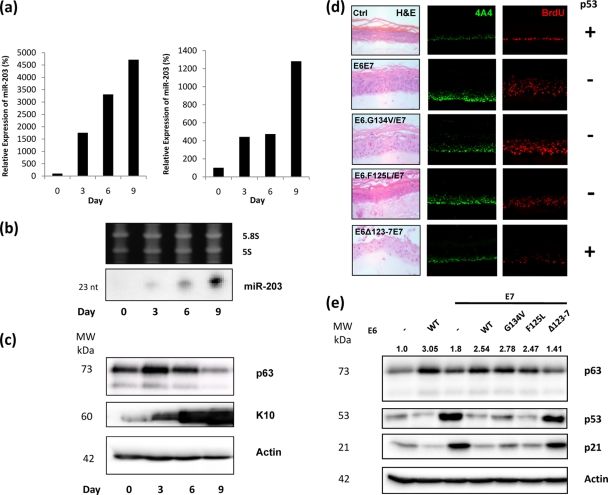
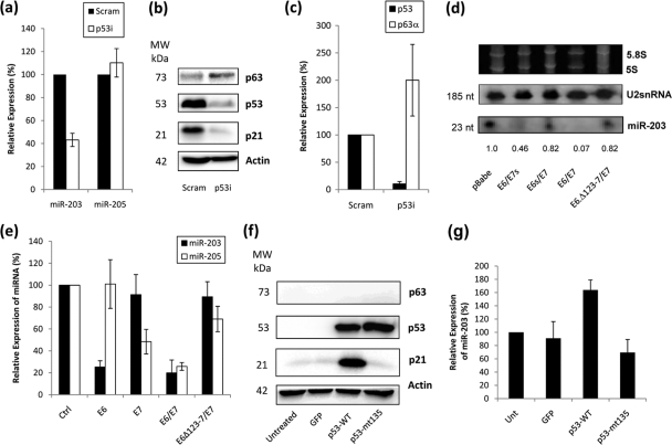
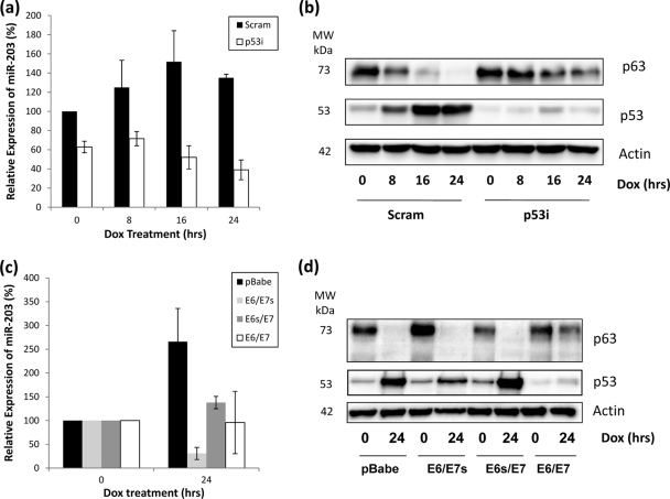
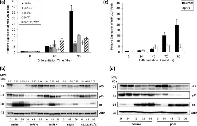
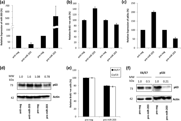
A screen of microRNA (miRNA) expression following differentiation in human foreskin keratinocytes (HFKs) identified changes in several miRNAs, including miRNA 203 (miR-203), which has previously been shown to play an important role in epithelial cell biology by regulating p63 levels. We investigated how expression of human papillomavirus type 16 (HPV16) oncoproteins E6 and E7 affected miR-203 expression during proliferation and differentiation of HFKs. We demonstrated that miR-203 expression is reduced in HFKs where p53 function is compromised, either by the viral oncoprotein E6 or by knockout of p53 using short hairpin RNAs (p53i). We show that the induction of miR-203 observed during calcium-induced differentiation of HFKs is significantly reduced in HFKs expressing E6 and in p53i HFKs. Induction of miR-203 in response to DNA damage is also reduced in the absence of p53. We report that proliferation of HFKs is dependent on the level of miR-203 expression and that overexpression of miR-203 can reduce overproliferation in E6/E7-expressing and p53i HFKs. In summary, these results indicate that expression of miR-203 is dependent on p53, which may explain how expression of HPV16 E6 can disrupt the balance between proliferation and differentiation, as well as the response to DNA damage, in keratinocytes.


Figures

FIG. 1.
FIG. 1.
p63 and miR-203 expression in organotypic rafts. (a) RQ-PCR analysis of RNA samples harvested from organotypic rafts at different time points. Results from two sets of raft samples, independently generated from separate HFK batches, demonstrate that miR-203 expression increases significantly over time. (b) This observation is confirmed by Northern blotting analysis. (c) Western blot analysis of matched protein samples demonstrates increased K10 expression, indicating that differentiation of HFKs is occurring in the rafts. p63 levels initially increase slightly and are then reduced. (d) p63 expression is dependent on the ability of E6 to degrade p53. p63 expression in Ctrl organotypic rafts is confined to basal cells (row 1). In rafts expressing E6 and E7, increased p63 expression is observed in upper layers of the raft, together with evidence of increased cell proliferation (row 2). This increase in both p63 expression and proliferation is also apparent in rafts expressing E7 and mutant E6 which can still degrade p53 (rows 3 and 4). However, in rafts expressing E7 and mutant E6 which cannot degrade p53, p63 expression and proliferation are similar to those for the control (row 5). (e) Western blot analysis confirms that p63 levels are significantly elevated in HFKs expressing E6 (lane 2), both E6 and E7 (lane 4), and both E6 mutants capable of degrading p53 and E7 (lanes 5 and 6). A more moderate increase is observed in HFKs expressing E7 alone (lane 3). HFKs expressing both E6 mutants incapable of degrading p53 and E7 show no increase in p63 expression (lane 7). Numbers above the p63 panel represent expression signals of p63 bands after normalization to actin expression. With the exception of panel a (n = 2), all images and blots are representative of three independent experiments performed on separate batches of HFKs.
FIG. 2.
FIG. 2.
miR-203 expression is p53 dependent. (a) RQ-PCR analysis demonstrates a reduction in miR-203 expression in HFKs when p53 was knocked down (p53i) compared to scramble controls (Scram). In comparison, the expression of miR-205, another highly expressed miRNA in the skin, did not change. (b) Western blot analysis confirms the knockdown of p53 in these cells and also demonstrates an increase in p63 expression. (c) RQ-PCR analysis confirms that knockdown of p53 at the mRNA level results in an increase in expression of p63α mRNA in these cells. (d) Northern blotting demonstrates that miR-203 expression is reduced in HFKs expressing E6 alone (lane 2) and both E6 and E7 (lane 4). In HFKs expressing E7 alone (lane 3) and E7 and mutant E6 (Δ123-7) (lane 5), which cannot degrade p53, miR-203 expression is only slightly decreased. Numbers below the panel represent the expression signal of miR-203 after normalization to the U2snRNA signal. (e) RQ-PCR analysis confirms that miR-203 expression is decreased in HFKs expressing E6 and both E6 and E7 compared to the control, but is not significantly reduced in HFKs expressing E7 alone and both E7 and mutant E6 (Δ123-7). In contrast, miR-205 levels are reduced in the presence of E7, regardless of the presence or function of E6. (f) Western blotting shows equal levels of wild-type and mutant p53 when expressed in a p53-null H1299 cell line, but upregulation of p21 is observed only with the former. Control experiments demonstrate that no expression of p53 is detected in untreated cells or cells transfected with a GFP-expressing vector. (g) Expression of wild-type p53 in H1299 cells results in an increase of miR-203 levels, as measured by RQ-PCR. In contrast, expression of dominant negative p53 mut135 does not induce miR-203, with expression levels similar to those for controls. All blots are representative of three independent experiments performed on separate batches of cells.
FIG. 3.
FIG. 3.
Induction of miR-203 following DNA damage is lost in p53i HFKs and HFKS expressing E6 and/or E7. (a) RQ-PCR shows that miR-203 expression increases following DNA damage induced by doxorubicin. However, this induction is not observed in p53i HFKs. (b) Western blotting demonstrates that p53 levels increase and p63 levels decrease in control cells after DNA damage. However, in the absence of p53 induction in p53i HFKs, this decrease in p63 levels is not as great, with detection still apparent at 24 h. (c) RQ-PCR shows that induction of miR-203 expression is not observed in HFKs expressing E6 alone (E6/E7s), E7 alone (E6s/E7), or both together (E6/E7) following DNA damage induced by doxorubicin. (d) Western blotting demonstrates that p53 levels increase and p63 levels decrease in control cells after DNA damage. A similar decrease is noted in E6/E7s and E6s/E7 HFKs, but higher residual p63 levels are apparent in E6/E7 HFKs after 24 h. All blots are representative of three independent experiments performed on different batches of primary HFKs.
FIG. 4.
FIG. 4.
Differentiation-specific upregulation of miR-203 expression is lost in E6/E7 and p53i HFKs. (a and c) RQ-PCR shows that the upregulation of miR-203 expression in control HFKs following calcium-induced differentiation is significantly reduced in HFKs expressing functional E6 and/or functional E7 (a) and p53i HFKs (c). RQ-PCR results are expressed relative to control values at T = zero. (b and d) Protein expression analysis from matched protein samples demonstrates that K1 induction is reduced in HFKs expressing E6 and/or E7, indicating lack of proper differentiation (b), but is not significantly altered in p53i HFKs (d). p63 levels are higher in HFKs expressing E6 and/or E7 than in controls, but reduction is not significantly affected in p53i HFKs compared to controls. Numbers above the p63 panel in panel b represent expression signals of p63 bands after normalization to actin expression. All blots are representative of three independent experiments performed on separate batches of HFKs.
FIG. 5.
FIG. 5.
Effects of miR-203 expression on HFK proliferation. (a) RQ-PCR demonstrating knockdown and overexpression of miR-203 in HFKs compared to the respective controls. (b) Proliferation, as measured by BrdU incorporation, is increased in HFKS in which miR-203 is inhibited and decreased in HFKs which overexpress miR-203. (c) RQ-PCR analysis showing that expression of p63α mRNA is increased in HFKs in which miR-203 is inhibited and decreased in HFKs which overexpress miR-203, relative to the respective controls, indicating that miR-203 levels control expression of the predominant isoform of p63 in HFKs. (d) Western blot confirming that protein levels of p63 are increased in HFKs in which miR-203 is inhibited and decreased in HFKs which overexpress miR-203, relative to the respective controls. Numbers above the panel represent expression signals of p63 bands after normalization to actin expression and are representative of three independent experiments performed on different batches of primary HFKs. (e) Proliferation, as measured by BrdU incorporation, is reduced in E6/E7 and p53i HFKs which overexpress miR-203. (f) Western blot demonstrating that levels of p63 are significantly reduced in E6/E7 and p53i HFKs which overexpress miR-203. Numbers above panels represent expression signal of p63 bands after normalization to actin expression and are representative of three independent experiments performed on different batches of primary HFKs.

Similar articles

Cited by

References

    1. Andl, T., E. P. Murchison, F. Liu, Y. Zhang, M. Yunta-Gonzalez, J. W. Tobias, C. D. Andl, J. T. Seykora, G. J. Hannon, and S. E. Millar. 2006. The miRNA-processing enzyme dicer is essential for the morphogenesis and maintenance of hair follicles. Curr. Biol. 16:1041-1049. - PMC - PubMed
    1. Bauersachs, J., and T. Thum. 2007. MicroRNAs in the broken heart. Eur. J. Clin. Invest. 37:829-833. - PubMed
    1. Bernstein, E., S. Y. Kim, M. A. Carmell, E. P. Murchison, H. Alcorn, M. Z. Li, A. A. Mills, S. J. Elledge, K. V. Anderson, and G. J. Hannon. 2003. Dicer is essential for mouse development. Nat. Genet. 35:215-217. - PubMed
    1. Bommer, G. T., I. Gerin, Y. Feng, A. J. Kaczorowski, R. Kuick, R. E. Love, Y. Zhai, T. J. Giordano, Z. S. Qin, B. B. Moore, O. A. MacDougald, K. R. Cho, and E. R. Fearon. 2007. p53-mediated activation of miRNA34 candidate tumor-suppressor genes. Curr. Biol. 17:1298-1307. - PubMed
    1. Brosh, R., R. Shalgi, A. Liran, G. Landan, K. Korotayev, G. H. Nguyen, E. Enerly, H. Johnsen, Y. Buganim, H. Solomon, I. Goldstein, S. Madar, N. Goldfinger, A. L. Børresen-Dale, D. Ginsberg, C. C. Harris, Y. Pilpel, M. Oren, and V. Rotter. 2008. p53-Repressed miRNAs are involved with E2F in a feed-forward loop promoting proliferation. Mol. Syst. Biol. 4:229. - PMC - PubMed

Publication types

MeSH terms