. 2010 Nov 25;116(22):4532-41.
doi: 10.1182/blood-2010-05-283309. Epub 2010 Aug 11.

The B-cell tumor-associated antigen ROR1 can be targeted with T cells modified to express a ROR1-specific chimeric antigen receptor

Affiliations

The B-cell tumor-associated antigen ROR1 can be targeted with T cells modified to express a ROR1-specific chimeric antigen receptor

Michael Hudecek et al. Blood. .

Abstract

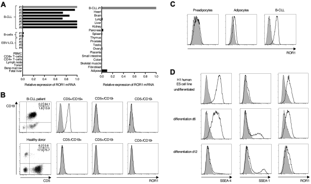
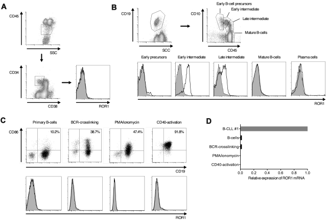
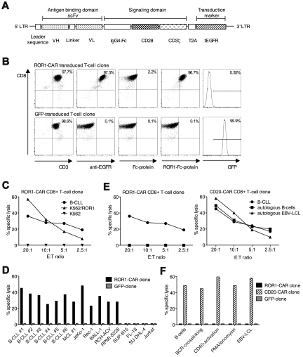
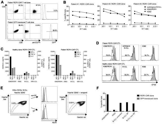
Monoclonal antibodies and T cells modified to express chimeric antigen receptors specific for B-cell lineage surface molecules such as CD20 exert antitumor activity in B-cell malignancies, but deplete normal B cells. The receptor tyrosine kinase-like orphan receptor 1 (ROR1) was identified as a highly expressed gene in B-cell chronic lymphocytic leukemia (B-CLL), but not normal B cells, suggesting it may serve as a tumor-specific target for therapy. We analyzed ROR1-expression in normal nonhematopoietic and hematopoietic cells including B-cell precursors, and in hematopoietic malignancies. ROR1 has characteristics of an oncofetal gene and is expressed in undifferentiated embryonic stem cells, B-CLL and mantle cell lymphoma, but not in major adult tissues apart from low levels in adipose tissue and at an early stage of B-cell development. We constructed a ROR1-specific chimeric antigen receptor that when expressed in T cells from healthy donors or CLL patients conferred specific recognition of primary B-CLL and mantle cell lymphoma, including rare drug effluxing chemotherapy resistant tumor cells that have been implicated in maintaining the malignancy, but not mature normal B cells. T-cell therapies targeting ROR1 may be effective in B-CLL and other ROR1-positive tumors. However, the expression of ROR1 on some normal tissues suggests the potential for toxi-city to subsets of normal cells.


Figures

Figure 1
Figure 1
ROR1 is uniformly expressed in B-CLL and shows expression in human ES cells. (A) ROR1 mRNA expression in B-CLL cells, normal primary and EBV-transformed B cells and a panel of normal hematopoietic and nonhematopoietic tissues was analyzed by qPCR. One B-CLL sample (B-CLL #1) was used as a reference (expression = 1), and the relative expression of ROR1 was compared with GAPDH as housekeeping gene. (B) Analysis of ROR1-protein expression on the cell surface of PBMCs obtained from B-CLL patients and normal donors using specific polyclonal antibodies (solid black line) compared with isotype controls (gray histogram). Data are representative for 10 B-CLL patients and 4 normal donors. (C) ROR1 expression on mature adipocytes that were generated in vitro by differentiation from human white preadipocytes, compared with B-CLL cells analyzed in the same experiment. (D) Expression of ROR1, SSEA-4, and SSEA-1 on the human ES cell line H1 before and after undirected in vitro differentiation for 6 and 12 days. Histograms show staining with specific monoclonal (SSEA-4/1) and polyclonal (ROR1) antibodies (solid black line) versus matched isotype control antibodies (gray histogram).
Figure 2
Figure 2
ROR1 is uniformly expressed on MCL. (A) Screening for expression of ROR1 on tumor cell lines and clinical samples of MCL (Jeko-1), multiple myeloma (RPMI-8226), and follicular lymphoma (FL-18). Data of clinical samples are representative for 6 MCL, 6 multiple myeloma, and 5 follicular lymphoma patients, respectively. (B) The B-cell precursor (B-ALL) tumor cell lines BALL-1 and RCH-ACV but not SUP-B15 are positive for ROR1-protein expression by flow cytometry. (C) The tumor cells lines Jurkat (T-cell acute lymphoblastic leu-kemia), SU-DHL-4 (diffuse large B-cell lymphoma), and K562 (chronic myeloid leukemia) are negative for ROR1 surface expression. Expression of ROR1 on K562 cells after stable transfection with the ROR1-gene (K562/ROR1).
Figure 3
Figure 3
ROR1 is expressed on a subset of normal B-cell precursors in adult bone marrow. (A-B) Multicolor flow cytometric analysis of BMMCs from healthy donors. Data are representative for results from 3 healthy donors. (A) Analysis of ROR1-expression on CD34+CD38lin hematopoietic progenitor cells. (B) Identification of B-cell precursors at distinct stages of B-cell development, mature B cells and plasma cells based on coexpression of CD19, CD10 and CD45, and analysis of ROR1-expression on the cell surface. Staining with specific polyclonal anti–human ROR1 antibodies (solid black line) versus isotype controls (gray histogram). (C) Activation of primary peripheral B cells of healthy donors by BCR-crosslinking, with PMA/ionomycin and stimulation through CD40. Expression of CD86 (top panel) and ROR1 (bottom panel) before and after activation. (D) ROR1 mRNA expression in normal resting, activated and EBV-transformed B cells analyzed by qPCR. The same B-CLL sample as in Figure 1A (B-CLL #1) was used as a reference and the relative expression of ROR1 compared with GAPDH as housekeeping gene.
Figure 4
Figure 4
A ROR1-specific CAR expressed in human CD8+ T cells confers specific recognition of ROR1-positive tumor cells. (A) Design of the transgene encoding the ROR1-CAR. The CAR-cassette contains a truncated extracellular EGFR transduction marker, separated by a cleavable T2A element to identify transduced T cells. (B) Phenotype and transgene expression of a ROR1-CAR transduced and a GFP-transduced CD8+ T-cell clone obtained from a healthy donor. To identify CAR-transduced T cells, primary staining was either performed with biotinylated anti-EGFR mAb or biotinylated recombinant Fc-ROR1 fusion protein that contained the extracellular domain of ROR1 and binds directly to the scFv of the CAR, and compared with staining with Fc-protein alone. Secondary staining was performed with streptavidin-conjugated PE. (C) Specific cytotoxicity of ROR1-CAR transduced CD8+ T cells against primary B-CLL and ROR1-transfected K562 cells by chromium release assay. Cytotoxicity data are presented as mean values of triplicate wells. The standard deviation of each triplicate was ≤ 3% in all cases (C-F). (D) ROR1-CAR modified CD8+ T cells but not T cells transduced with a GFP-encoding control vector specifically recognize primary B-CLL and MCL samples from multiple patients and a panel of ROR1-positive tumor cell lines. Cytotoxicity was analyzed by chromium release assay at an E/T ratio of 20:1. (E) Chromium release assay comparing the cytotoxicity of ROR1-CAR and CD20-CAR transduced CD8+ T cells obtained from the same donor against primary B-CLL cells and autologous resting and EBV-transformed B cells. (F) Recognition of autologous B cells that had been activated by BCR-crosslinking, with PMA/ionomycin, and stimulation through CD40 by ROR1-CAR, CD20-CAR, and control GFP-transduced CD8+ T cells by chromium release assay at an E/T ratio of 20:1.
Figure 5
Figure 5
ROR1-CAR transduced CD8+ T cells obtained from B-CLL patients specifically recognize autologous tumor cells. (A) Phenotype and transgene expression of a ROR1-CAR and a GFP-transduced CD8+ T-cell clone obtained from a B-CLL patient. Data are representative for results obtained in 3 B-CLL patients. (B) ROR-CAR CD8+ T cells from 3 B-CLL patients specifically recognize autologous primary B-CLL and ROR1-positive tumor cells by chromium release assay. Cytotoxicity data are presented as mean values of triplicate wells. The standard deviation of each triplicate was ≤ 3% in all cases (B,F). (C) Secretion of INF-γ (left axis), TNF-α and IL-2 (right axis) of ROR1-CAR transduced CD8+ T cells obtained from a healthy donor and a B-CLL patient in response to primary (autologous) B-CLL and ROR1-positive tumor cell lines after incubation for 24 hours at an E:T ratio of 2:1 quantified by Luminex Assay. (D) Proliferation of ROR1-CAR expressing CD8+ T cells obtained from a healthy donor and a B-CLL patient after stimulation with primary (autologous) B-CLL and ROR1-positive tumor cell lines assessed by CFSE dilution after incubation for 72 hours at an E/T ratio of 2:1. Specific proliferation (black solid line) versus background proliferation (gray histogram). (E) Detection of a side population of Hoechst 33342 effluxing B-CLL cells by flow cytometry after staining of primary B-CLL cells with Hoechst 33342 (left dot plot). Expression of ROR1 on SP and non-SP B-CLL cells (center histograms). Inhibition of Hoechst 33342 efflux by addition of 50μM verapamil (right dot plot). Data are representative for results obtained in 3 B-CLL patients. (F) Specific and equivalent elimination of sort-purified SP and non-SP primary B-CLL cells by ROR1-CAR transduced CD8+ T cells by chromium release assay at an E/T ratio of 20:1.

Comment in

  • RORing T cells target CLL and MCL.
    Sadelain M. Sadelain M. Blood. 2010 Nov 25;116(22):4387-8. doi: 10.1182/blood-2010-09-303388. Blood. 2010. PMID: 21109622 No abstract available.

References

    1. Sorror ML, Storer BE, Sandmaier BM, et al. Five-year follow-up of patients with advanced chronic lymphocytic leukemia treated with allogeneic hematopoietic cell transplantation after nonmyeloablative conditioning. J Clin Oncol. 2008;26(30):4912–4920. - PMC - PubMed
    1. Maris MB, Sandmaier BM, Storer BE, et al. Allogeneic hematopoietic cell transplantation after fludarabine and 2 Gy total body irradiation for relapsed and refractory mantle cell lymphoma. Blood. 2004;104(12):3535–3542. - PubMed
    1. Nishida T, Hudecek M, Kostic A, et al. Development of tumor-reactive T cells after nonmyeloablative allogeneic hematopoietic stem cell transplant for chronic lymphocytic leukemia. Clin Cancer Res. 2009;15(14):4759–4768. - PMC - PubMed
    1. Hudecek M, Anderson LJ, Nishida T, Riddell S. Adoptive T-cell therapy for B-cell malignancies. Expert Rev Hematol. 2009;2(5):517–532. - PMC - PubMed
    1. Maher J, Brentjens RJ, Gunset G, Riviere I, Sadelain M. Human T-lymphocyte cytotoxicity and proliferation directed by a single chimeric TCRzeta /CD28 receptor. Nat Biotechnol. 2002;20(1):70–75. - PubMed

Publication types

MeSH terms

Substances