Comparative Study
. 2011 Jan 21;52(1):339-44.
doi: 10.1167/iovs.10-5944. Print 2011 Jan.

Clinical and antiviral efficacy of an ophthalmic formulation of dexamethasone povidone-iodine in a rabbit model of adenoviral keratoconjunctivitis

Affiliations
Comparative Study

Clinical and antiviral efficacy of an ophthalmic formulation of dexamethasone povidone-iodine in a rabbit model of adenoviral keratoconjunctivitis

Christian Clement et al. Invest Ophthalmol Vis Sci. .

Abstract

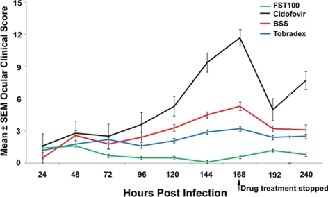
Purpose: To determine the efficacy of a new formulation of topical dexamethasone 0.1%/povidone-iodine 0.4% (FST-100) in reducing clinical symptoms and infectious viral titers in a rabbit model of adenoviral keratoconjunctivitis.

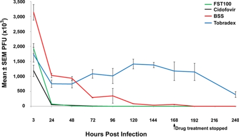
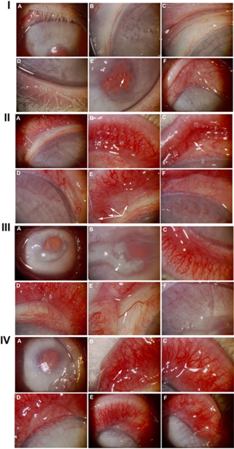
Methods: Rabbit corneas were inoculated bilaterally with 2×10(6) plaque-forming-units (PFU) of adenovirus type 5 (Ad5) after corneal scarification. Animals were randomized 1:1:1:1 (five rabbits per group) to FST-100, 0.5% cidofovir, tobramycin/dexamethasone (Tobradex; Alcon Laboratories, Fort Worth, TX) ophthalmic suspension, and balanced salt solution (BSS; Alcon Laboratories). Treatment began 12 hours after viral inoculation and continued for 7 consecutive days. The eyes were clinically scored daily for scleral inflammation (injection), ocular neovascularization, eyelid inflammation (redness), friability of vasculature, inflammatory discharge (pus), and epiphora (excessive tearing). Eye swabs were collected daily before treatment for the duration of the study. Virus was eluted from the swabs and PFU determined by titration on human A549 cells, according to standard procedures.

Results: The FST-100 treatment resulted in significantly lower clinical scores (P<0.05) than did the other treatments. The 0.5% cidofovir exhibited the most ocular toxicity compared with FST-100, tobramycin/dexamethasone, and balanced salt solution treatments. FST-100 and 0.5% cidofovir significantly (P<0.05) reduced viral titers compared with tobramycin/dexamethasone or balanced salt solution.

Conclusions: FST-100 was the most efficacious in minimizing the clinical symptoms of adenovirus infection in rabbit eyes. FST-100 and 0.5% cidofovir were both equally effective in reducing viral titers and decreasing the duration of viral shedding. By providing symptomatic relief in addition to reducing infectious virus titers, FST-100 should be a valuable addition to treatment of epidemic adenoviral keratoconjunctivitis.


Figures

Figure 1.
Figure 1.
Line graph showing mean ± SEM ocular clinical scores of Ad5-infected rabbit eyes treated with FST-100, cidofovir, balanced salt solution (BSS; Alcon Laboratories), or 0.3% tobramycin/0.1% dexamethasone (Tobradex; Alcon Laboratories) over time. The mean ± SEM ocular clinical score is calculated from the daily cumulative of the six clinical parameters (scleral inflammation [injection], ocular neovascularization, eyelid inflammation [redness], friability of vasculature, inflammatory discharge [pus], and epiphora [excessive tearing]) per rabbit eye, indicating that a normal, uninfected eye would have a score of 0 and an eye with the most severe disease would have a score of 18.
Figure 2.
Figure 2.
Line graph showing mean ± SEM PFU reduction by FST-100, cidofovir, balanced salt solution (BSS; Alcon Laboratories), and 0.3% tobramycin/0.1% dexamethasone (Tobradex; Alcon Laboratories). When the SEM error bars are not visible, they are too small to be seen. Swabs from infected eyes were eluted and 200 μL of 10-fold serial dilutions of the original eluate/swab plated on near-confluent A549 cells with methylcellulose overlay. Fixed cells were stained with Hucker's modified crystal violet solution after 3 days and the plaques counted.
Figure 3.
Figure 3.
(I) Image of FST-100–treated Ad5-infected NZW rabbit eyes at postinfection day 7 (168 hours). (IA) Intact cornea; (IB) no prominent blood vessels on cornea; (IC) upper eyelid with no inflammation; (ID) lower eyelid with no inflammation; (IE) lower eyelid with no inflammation horizontal to the clearly visible cross-hatch virus inoculation scratch (arrow) and (IF) no active blood vessel formation on the cornea. (II) Image of 0.5% cidofovir–treated Ad5-infected NZW rabbit eyes at postinfection day 7 (168 hours). (IIA) Intact cornea with inflammation and injection; (IIB) upper eyelid inflammation, injection with friable neovascularization; (IIC) upper eyelid inflammation with subconjunctival heme (arrow); (IID) neovascularization with active blood vessels (appearing to protrude from the limbus) forming on the cornea; (IIE) prominent blood vessels (not appearing to protrude from limbus, arrows) spreading onto the cornea; (IIF) network of blood vessels developing on the cornea. (III) Image of balanced salt solution–treated Ad5-infected NZW rabbit eyes at postinfection day 7 (168 hours). (IIIA) Intact cornea; (IIIB) discharge and exudates on the cornea (arrowhead); (IIIC) lower eyelid inflammation, injection with friable neovascularization; (IIID) upper eyelid inflammation, injection with friable neovascularization; (IIIE) active blood vessel network on the eyelid margins and prominent blood vessel (arrow) close to the caruncle of the eye; (IIIF) meshwork of small blood vessels on the cornea. (IV) Image of 0.3% tobramycin/0.1% dexamethasone–treated Ad5-infected NZW rabbit eyes at postinfection day 7 (168 hours). (IVA) Intact cornea; (IVB) upper eyelid inflammation with subconjunctival heme (arrow); (IVC) upper eyelid inflammation, injection with friable neovascularization; (IVD) neovascularization with active blood vessel formation on the cornea; (IVE) upper eyelid inflammation, injection with friable neovascularization; (IVF) upper eyelid with friable neovascularization, blood vessel formation on the eyelid margins, and neovascularization.

References

    1. De Jong JC, Wermenbol AG, Verweij-Uijterwaal MW, et al. Adenoviruses from human immunodeficiency virus-infected individuals, including two strains that represent new candidate serotypes Ad50 and Ad51 of species B1 and D, respectively. J Clin Microbiol. 1999;37:3940–3945 - PMC - PubMed
    1. Schrauder A, Altmann D, Laude G, et al. Epidemic conjunctivitis in Germany. 2004. Eur Surveill. 2006;11:185–187 - PubMed
    1. Asencio-Durán M, Romero-Martín R, García-Martínez JR, et al. Nosocomial outbreak of epidemic keratoconjunctivitis in a neonatal intensive care unit (in Spanish). Arch Soc Esp Oftalmol. 2007;82:73–79 - PubMed
    1. Kumar NL, Black D, McClellan K. Daytime presentations to a metropolitan ophthalmic emergency department. Clin Exp Ophthalmol. 2005;33:586–592 - PubMed
    1. Ford E, Nelson KE, Warren D. Epidemiology of epidemic keratoconjunctivitis. Epidemiol Rev. 1987;9:244–261 - PubMed

Publication types

MeSH terms