. 2010 Nov;51(11):3259-69.
doi: 10.1194/jlr.M009365. Epub 2010 Aug 11.

Biological activities of 7-dehydrocholesterol-derived oxysterols: implications for Smith-Lemli-Opitz syndrome

Affiliations

Biological activities of 7-dehydrocholesterol-derived oxysterols: implications for Smith-Lemli-Opitz syndrome

Zeljka Korade et al. J Lipid Res. 2010 Nov.

Abstract

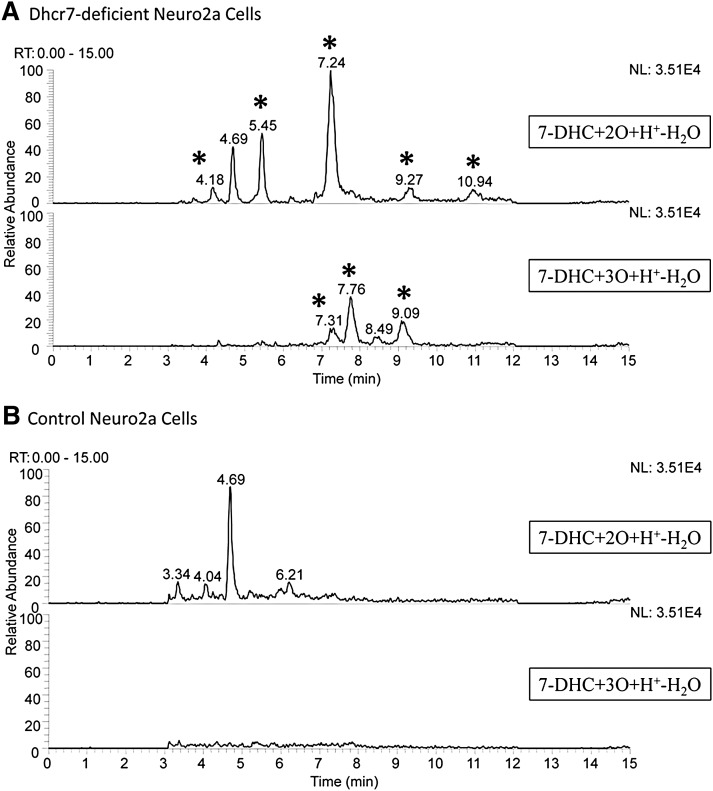
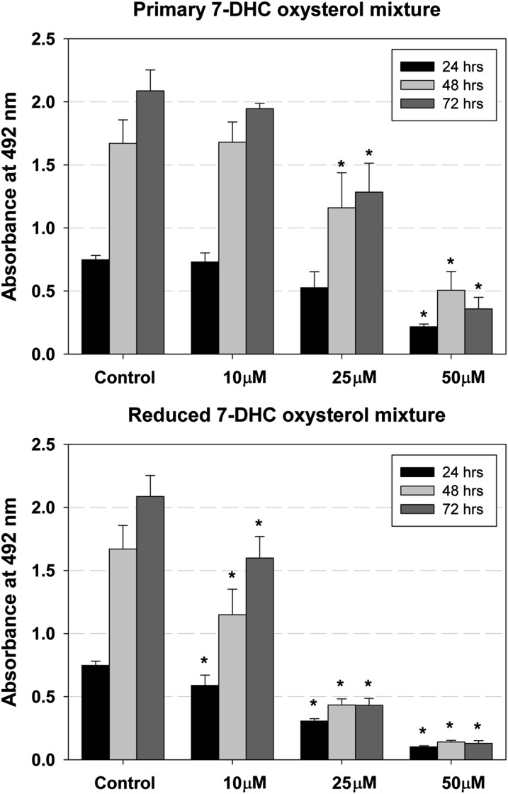
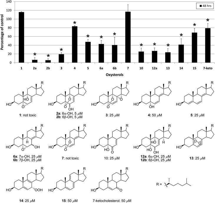
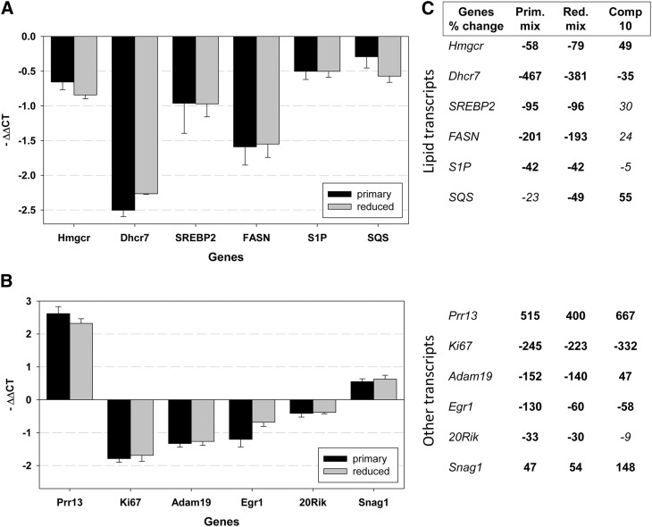
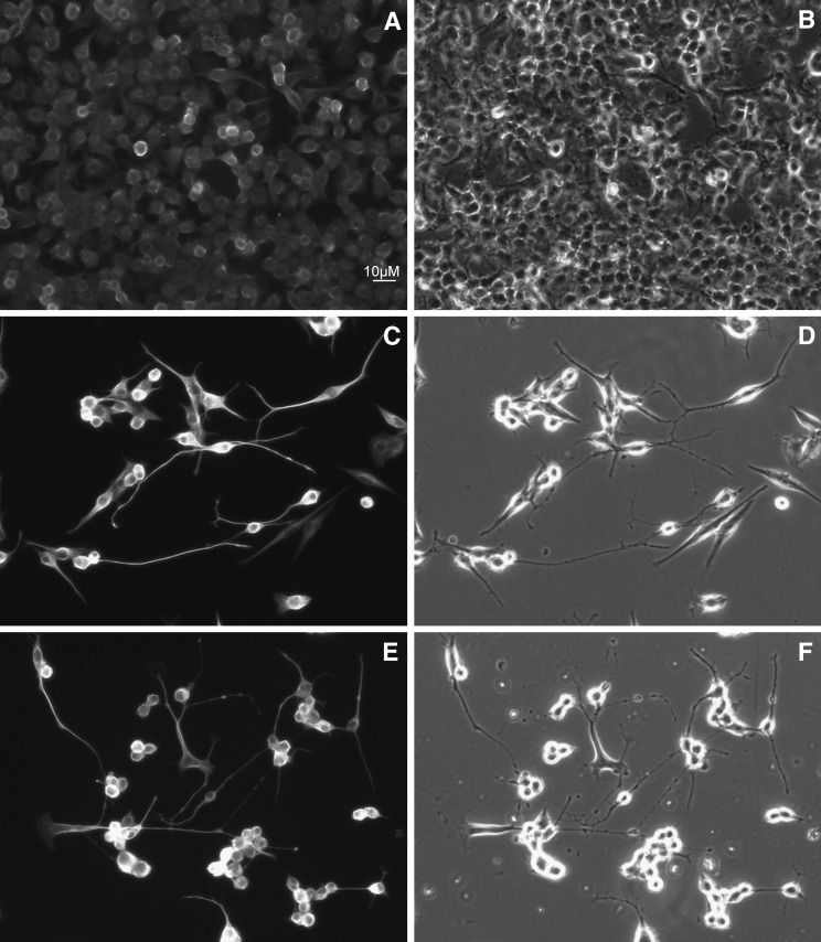
Smith-Lemli-Opitz syndrome (SLOS) is a metabolic and developmental disorder caused by mutations in the gene encoding the enzyme 7-dehydrocholesterol reductase (Dhcr7). This reductase catalyzes the last step in cholesterol biosynthesis, and levels of 7-dehydrocholesterol (7-DHC), the substrate for this enzyme, are elevated in SLOS patients as a result of this defect. Our group has previously shown that 7-DHC is extremely prone to free radical autoxidation, and we identified about a dozen different oxysterols formed from oxidation of 7-DHC. We report here that 7-DHC-derived oxysterols reduce cell viability in a dose- and time-dependent manner, some of the compounds showing activity at sub-micromolar concentrations. The reduction of cell survival is caused by a combination of reduced proliferation and induced differentiation of the Neuro2a cells. The complex 7-DHC oxysterol mixture added to control Neuro2a cells also triggers the gene expression changes that were previously identified in Dhcr7-deficient Neuro2a cells. Based on the identification of overlapping gene expression changes in Dhcr7-deficient and 7-DHC oxysterol-treated Neuro2a cells, we hypothesize that some of the pathophysiological findings in the mouse SLOS model and SLOS patients might be due to accumulated 7-DHC oxysterols.


Figures

Fig. 1.
Fig. 1.
7-DHC oxysterols are present in Dhcr7-deficient Neuro2a cells. The chromatogram in panel A shows the oxysterol profile of Dhcr7-deficient Neuro2a cells. New peaks relative to control (B) are marked with an asterisk. The peak at RT = 4.69 min represents a minor MS fragment from the external standard 25-hydroxycholesterol.
Fig. 2.
Fig. 2.
Primary and reduced 7-DHC oxysterol mixtures reduce cell viability in a dose- and time-dependent manner. The x-axis shows different concentrations of oxysterol mixture, the y-axis shows absorbance at 492 nm, and different colors of the bars show different time points. Under control conditions, the number of cultured cells increases over the 72 h time period. Different concentrations of the 7-DHC oxysterol mixture have specific effects on cell survival; whereas treatment with 10 μM concentration of primary mixture does not differ from control, 10 μM of the reduced mixture significantly reduces the cell number. The error bars show SD and the stars show significant change (P < 0.01).
Fig. 3.
Fig. 3.
Individual 7-DHC oxysterols show toxicity equal to or greater than the oxysterol mixture, correlation of structures with cellular effects. A: Neuro2a cells were treated with purified oxysterols (25 µM), and cell survival was analyzed 48 h later. At this concentration, a majority of compounds reduces cell viability to <50% (P < 0.001, marked with star). Compounds 1 and 7 are not toxic and compound 4 reduces cell survival by 20% (P < 0.01). B: Chemical structures of oxysterols. Compounds 1–12 are formed from free radical oxidation of 7-DHC. Compounds 13–15 are formed from photo-oxidation of 7-DHC. The number below each compound shows the oxysterol concentration that leads to reduction in cell survival to ≤50% at 48 h after the treatment.
Fig. 4.
Fig. 4.
Oxysterol treatment of control Neuro2a leads to gene expression changes. In the graphs A and B, the genes are plotted on the x-axis, and the y-axis denotes the average –ΔΔCt that was calculated against Tbp1 as a normalizer. P < 0.001 for changes shown in bold and not significant for those in Italic. A: Oxysterol treatment results in decreased expression of critical lipid biosynthesis genes that were also found altered in Dhcr7-deficient Neuro2a cells. The results are concordant for the primary (50 µM) and reduced (25 µM) mixtures. Compound 10 significantly affects the expression of Dhcr7 at both 24 and 48 h, but the effect on other lipid genes is not consistent and not significant. B: Oxysterol treatment results in gene expression changes similar to those identified in Dhcr7-deficient Neuro2a cells. Whereas Prr13, Egr1, and Snag1 showed consistent changes at both the 24 and 48 h time points, Adam19 and 20Rik were not changed at 48 h, suggesting transient change in the expression of these genes in response to oxysterol treatment. Compound 10 affects the expression of Ki67, Prr13, Egr1, and Snag1 similarly to primary and reduced mixtures. C: Summary of gene expression changes expressed as percentage of untreated cells. The table lists the genes and percentage of change. The changes are upregulation (positive value) and downregulation (negative value). For details of gene expression changes in Dhcr7-deficient cells, see Korade et al. (40).
Fig. 5.
Fig. 5.
Both primary and reduced 7-DHC oxysterol mixtures induce differentiation of Neuro2a cells. After 48 h of oxysterol treatment, cells were fixed and processed for immunocytochemistry using Tu20 (A, C, E) antibody. Panels on the left show Cy3 fluorescence and the panels on the right show phase contrast of the same field. A and B are control, C and D are treated with 50 μM primary 7-DHC oxysterol mixture, and E and F are treated with 25 μM reduced mixture.

References

    1. Porter N. A. 1986. Mechanisms for the autoxidation of polyunsaturated lipids. Acc. Chem. Res. 19: 262–270.
    1. Porter N. A., Caldwell S. E., Mills K. A. 1995. Mechanisms of free radical oxidation of unsaturated lipids. Lipids. 30: 277–290. - PubMed
    1. Yin H., Porter N. A. 2005. New insights regarding the autoxidation of polyunsaturated fatty acids. Antioxid. Redox Signal. 7: 170–184. - PubMed
    1. Porter N. A., Funk M. O., Gilmore D. W., Isaac R., Nixon J. 1976. The formation of cyclic peroxides from unsaturated hydroperoxides: models for prostaglandin biosynthesis. J. Am. Chem. Soc. 98: 6000–6005. - PubMed
    1. Porter N. A., Weber B. A., Weenen H., Khan J. A. 1980. Autoxidation of polyunsaturated lipids. Factors controlling the stereochemistry of product hydroperoxides. J. Am. Chem. Soc. 102: 5597–5601.

Publication types

MeSH terms

Substances