

Functional redundancy of Langerhans cells and Langerin+ dermal dendritic cells in contact hypersensitivity
- PMID: 20703247
- PMCID: PMC2980552
- DOI: 10.1038/jid.2010.223
Functional redundancy of Langerhans cells and Langerin+ dermal dendritic cells in contact hypersensitivity
Abstract
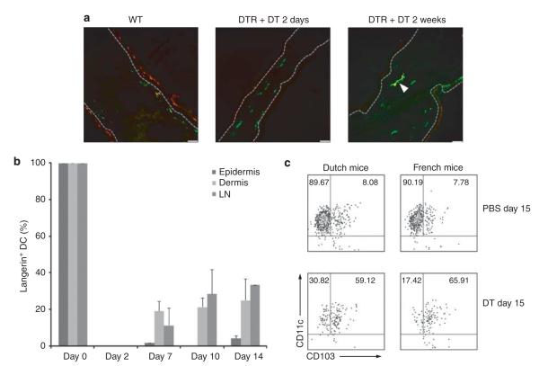
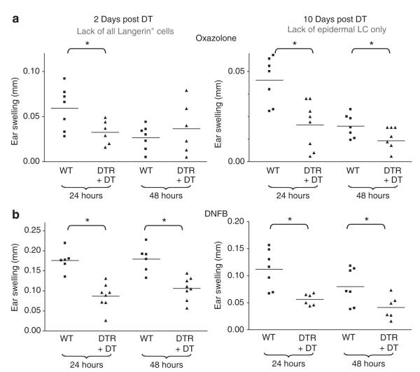
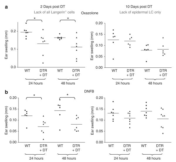
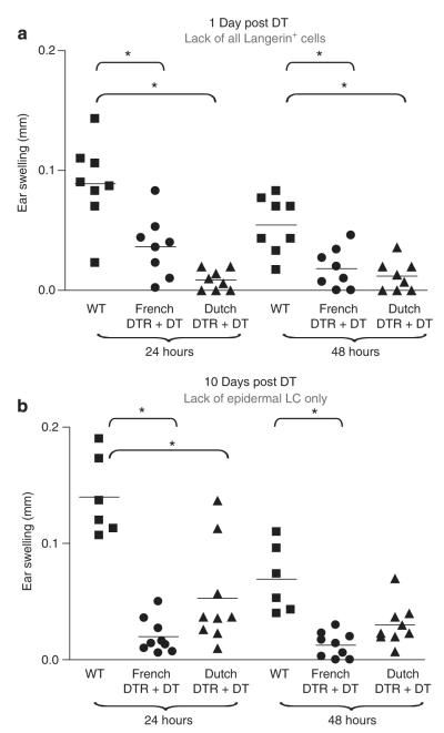
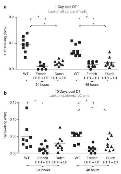
The relative roles of Langerhans cells (LC), dermal dendritic cells (DC), and, in particular, the recently discovered Langerin(+) dermal DC subset in the induction and control of contact hypersensitivity (CHS) responses remain controversial. Using an inducible mouse model, in which LC and other Langerin(+) DC can be depleted by injection of diphtheria toxin, we previously reported impaired transport of topically applied antigen to draining lymph nodes and reduced CHS in the absence of all Langerin(+) skin DC. In this study, we demonstrate that mice with a selective depletion of LC exhibit attenuated CHS only upon sensitization with a low hapten dose but not with a high hapten dose. In contrast, when painting a higher concentration of hapten onto the skin, which leads to increased antigen dissemination into the dermis, CHS is still diminished in mice lacking all Langerin(+) skin DC. Taken together, these data suggest that the magnitude of a CHS reaction depends on the number of skin DC, which have access to the hapten, rather than on the presence or absence of a particular skin DC population. LC and (Langerin(+)) dermal DC thus seem to have a redundant function in regulating CHS.
Figures
Similar articles
-
Inducible ablation of mouse Langerhans cells diminishes but fails to abrogate contact hypersensitivity.J Cell Biol. 2005 May 23;169(4):569-76. doi: 10.1083/jcb.200501071. Epub 2005 May 16. J Cell Biol. 2005. PMID: 15897263 Free PMC article.
-
Acute ablation of Langerhans cells enhances skin immune responses.J Immunol. 2010 Oct 15;185(8):4724-8. doi: 10.4049/jimmunol.1001802. Epub 2010 Sep 20. J Immunol. 2010. PMID: 20855870 Free PMC article.
-
Conditional deletion of TGF-βR1 using Langerin-Cre mice results in Langerhans cell deficiency and reduced contact hypersensitivity.J Immunol. 2011 Nov 15;187(10):5069-76. doi: 10.4049/jimmunol.1101880. Epub 2011 Oct 12. J Immunol. 2011. PMID: 21998450
-
Insights into Langerhans cell function from Langerhans cell ablation models.Eur J Immunol. 2008 Sep;38(9):2369-76. doi: 10.1002/eji.200838397. Eur J Immunol. 2008. PMID: 18792030 Review.
-
Langerhans cells and more: langerin-expressing dendritic cell subsets in the skin.Immunol Rev. 2010 Mar;234(1):120-41. doi: 10.1111/j.0105-2896.2009.00886.x. Immunol Rev. 2010. PMID: 20193016 Free PMC article. Review.
Cited by
-
Langerhans cell antigen capture through tight junctions confers preemptive immunity in experimental staphylococcal scalded skin syndrome.J Exp Med. 2011 Dec 19;208(13):2607-13. doi: 10.1084/jem.20111718. Epub 2011 Dec 5. J Exp Med. 2011. PMID: 22143886 Free PMC article.
-
Fate mapping analysis reveals a novel murine dermal migratory Langerhans-like cell population.Elife. 2021 Mar 26;10:e65412. doi: 10.7554/eLife.65412. Elife. 2021. PMID: 33769279 Free PMC article.
-
Cutting edge: identification of the thymic stromal lymphopoietin-responsive dendritic cell subset critical for initiation of type 2 contact hypersensitivity.J Immunol. 2013 Nov 15;191(10):4903-7. doi: 10.4049/jimmunol.1302175. Epub 2013 Oct 11. J Immunol. 2013. PMID: 24123684 Free PMC article.
-
Oral administration of lipoteichoic acid from Lactobacillus rhamnosus GG overcomes UVB-induced immunosuppression and impairs skin tumor growth in mice.Eur J Immunol. 2019 Nov;49(11):2095-2102. doi: 10.1002/eji.201848024. Epub 2019 Jul 31. Eur J Immunol. 2019. PMID: 31334839 Free PMC article.
-
Langerin+CD8+ Dendritic Cells in the Splenic Marginal Zone: Not So Marginal After All.Front Immunol. 2019 Apr 12;10:741. doi: 10.3389/fimmu.2019.00741. eCollection 2019. Front Immunol. 2019. PMID: 31031751 Free PMC article. Review.
References
-
- Bacci S, Alard P, Dai R, et al. High and low doses of haptens dictate whether dermal or epidermal antigen-presenting cells promote contact hypersensitivity. Eur J Immunol. 1997;27:442–8. - PubMed
-
- Banchereau J, Briere F, Caux C, et al. Immunobiology of dendritic cells. Annu Rev Immunol. 2000;18:767–811. - PubMed
-
- Banchereau J, Steinman RM. Dendritic cells and the control of immunity. Nature. 1998;392:245–52. - PubMed
-
- Bennett CL, Clausen BE. DC ablation in mice: promises, pitfalls, and challenges. Trends Immunol. 2007;28:525–31. - PubMed
-
- Bennett CL, Noordegraaf M, Martina CA, et al. Langerhans cells are required for efficient presentation of topically applied hapten to T cells. J Immunol. 2007;179:6830–5. - PubMed
Publication types
MeSH terms
Substances
Grants and funding
LinkOut - more resources
Full Text Sources
Other Literature Sources
Molecular Biology Databases
