. 2010 Aug 12;466(7308):829-34.
doi: 10.1038/nature09262.

Mesenchymal and haematopoietic stem cells form a unique bone marrow niche

Affiliations

Mesenchymal and haematopoietic stem cells form a unique bone marrow niche

Simón Méndez-Ferrer et al. Nature. .

Abstract

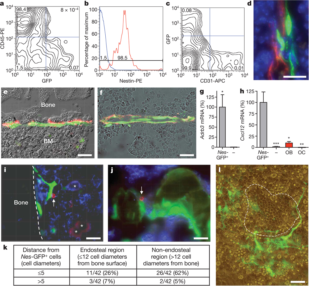
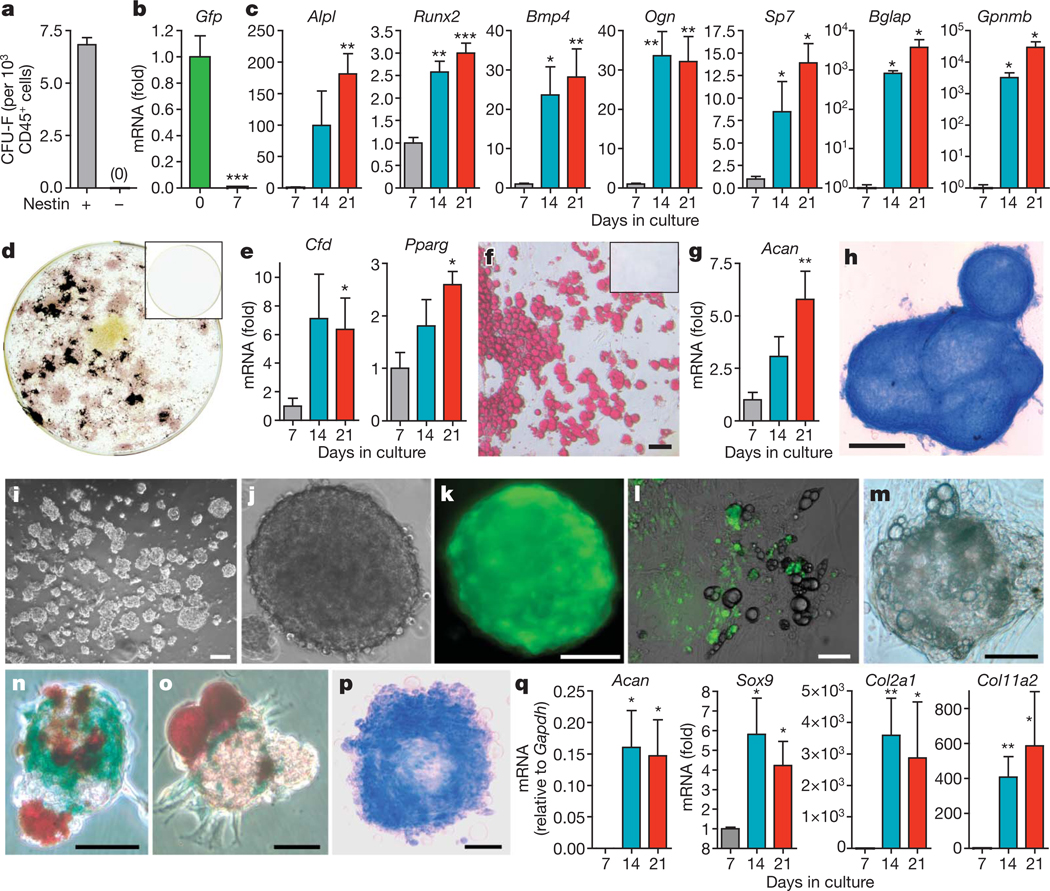
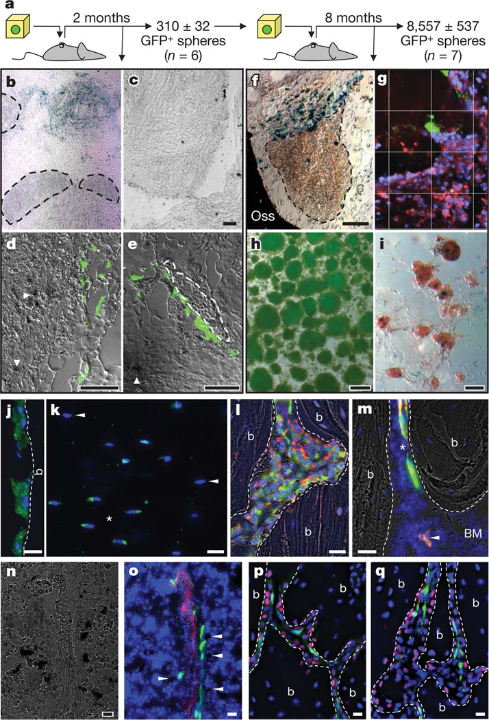
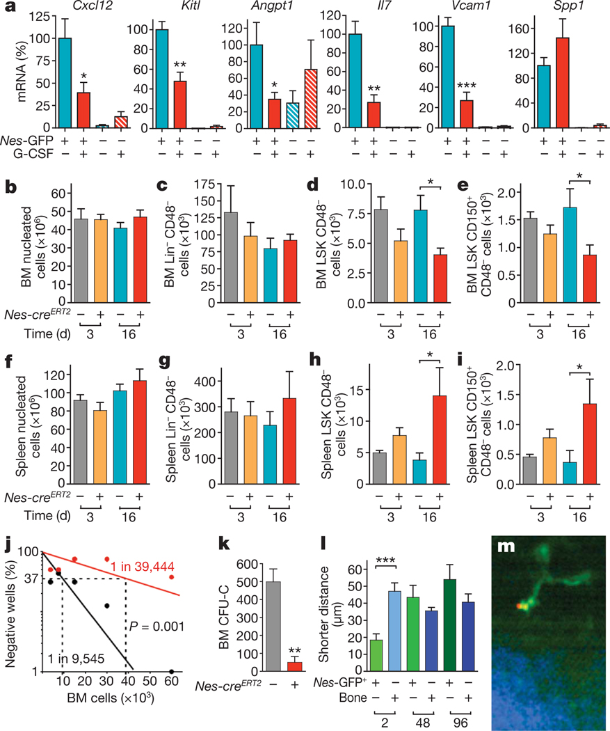
The cellular constituents forming the haematopoietic stem cell (HSC) niche in the bone marrow are unclear, with studies implicating osteoblasts, endothelial and perivascular cells. Here we demonstrate that mesenchymal stem cells (MSCs), identified using nestin expression, constitute an essential HSC niche component. Nestin(+) MSCs contain all the bone-marrow colony-forming-unit fibroblastic activity and can be propagated as non-adherent 'mesenspheres' that can self-renew and expand in serial transplantations. Nestin(+) MSCs are spatially associated with HSCs and adrenergic nerve fibres, and highly express HSC maintenance genes. These genes, and others triggering osteoblastic differentiation, are selectively downregulated during enforced HSC mobilization or beta3 adrenoreceptor activation. Whereas parathormone administration doubles the number of bone marrow nestin(+) cells and favours their osteoblastic differentiation, in vivo nestin(+) cell depletion rapidly reduces HSC content in the bone marrow. Purified HSCs home near nestin(+) MSCs in the bone marrow of lethally irradiated mice, whereas in vivo nestin(+) cell depletion significantly reduces bone marrow homing of haematopoietic progenitors. These results uncover an unprecedented partnership between two distinct somatic stem-cell types and are indicative of a unique niche in the bone marrow made of heterotypic stem-cell pairs.


Figures

Figure 1
Figure 1. Nes-GFP+ cells are perivascular stromal cells targeted by the SNS, express high levels of Cxcl12 and are physically associated with HSCs
ac, Flow cytometry characterization of bone marrow Nes-GFP+ cells; representative diagrams were obtained from bone marrow nucleated cells (a, c) or GFP+ cells (b); the percentage of the population in each quadrant is indicated. d, Projection image from ~10 µm Z-stack showing fluorescent signals from Nes-GFP (green), CD31+ vascular endothelial cells (red) and nuclei counterstained with DAPI (blue). e, f, Nes-GFP+ cells are directly innervated by tyrosine hydroxylase+ (red) catecholaminergic fibres in the endosteum (e) and the bone marrow parenchyma (f). g, h, Q-PCR from the same number of sorted CD45 Nes-GFP+ and CD45 Nes-GFP cells, and from primary osteoblasts (OB) and osteoclasts (OC; n = 5–10); *P < 0.05, **P < 0.01, ***P < 0.001; unpaired two-tailed t-test. Error bars indicate s.e.m. i, j, Immunostaining for CD150 (red), CD48 and haematopoietic lineages (blue) in femoral sections of Nes-Gfp mice. Stacked images of CD150+CD48Lin cells (arrow) adjacent to Nes-GFP+ cells in the endosteum (i) and the sinusoids (j). i, CD150+CD48+/Lin+ megakaryocytes (asterisks). k, Localization and number of CD150+CD48Lin cells relative to Nes-GFP+ cells in different bone marrow regions. l, Nes-GFP+ cells (green) and cobblestone-forming area (dashed line) in 4-week primary myeloid culture. e, f, l, Overlapped fluorescence and phase contrast images. All scale bars, 50 µm.
Figure 2
Figure 2. Nes-GFP+ cells are mesenchymal stem cells
a, Number of colony-forming units-fibroblast (CFU-F) in sorted CD45 Nes-GFP+ and CD45 Nes-GFP cells (n = 12). bh, Differentiation of sorted CD45 Nes-GFP+ cells (n = 4). Q-PCR of Gfp and (b) genes associated with osteoblastic (c; alkaline phosphatise (Alpl), Runx2, bone morphogenetic protein-4 (Bmp4), osteoglycin (Ogn), osterix (Sp7), osteocalcin (Bglap), osteoactivin (Gpnmb)), adipogenic (e; adipsin (Cfd), peroxisome proliferator-activated receptor gamma 2 (Pparg)) and chondrogenic (g; aggrecan (Acan)) differentiation is shown. d, f, h, Fully differentiated phenotypes of sorted CD45 Nes-GFP+ cells shown by alkaline phosphatase (pink) and Von Kossa (black) staining (d), Oil redO (red) staining (f) and Alcian blue (blue) staining (h). CD45Nes-GFP cells did not generate any progeny (d, f; inset). iq, Nes-GFP+ cells, but not the remaining CD45 bone marrow population, formed self-renewing and multipotent clonal spheres after 7–10 days in low-density culture. l, m, Adherent bulk-cultured CD45 Nes-GFP+ cells (l) or clonal spheres (m) lost GFP expression and differentiated into GFP adipocytes (refringent lipoid droplets). n, o, Representative mesenspheres showing spontaneous multilineage differentiation into Col2.3-LacZ+ osteoblasts (blue) and Oil red O+ adipocytes (red). p, q, Chondrogenic differentiation of mesenspheres shown by Alcian blue staining (p; blue) and Q-PCR for sex determining region Y-box 9 (Sox9), aggrecan (Acan), collagen type IIα1 (Col2α1) and collagen type XIα2 (Col11α2) (n = 4–11) (q). i, j, m, p, Bright field; k, Fluorescence. l, Overlapped fluorescence and bright field. Scale bars: 100 µm (f, l, k, n, p); 500 µm (h); 50 µm (l, m, o). Asterisks, where indicated, are: *P < 0.05; **P < 0.01; ***P < 0.001; unpaired two-tailed t-test. All error bars indicate s.e.m.
Figure 3
Figure 3. Adult nestin+ MSCs self-renew, differentiate and transfer haematopoietic activity in vivo
ai, In vivo self-renewal of adult bone marrow CD45 Nes-GFP+ cells in secondary transplants. a, Scheme showing the experimental paradigm. be, Primary ossicles showing numerous β-galactosidase+ osteoblasts derived from CD45 Nes-GFP+ cells (b, blue; d, e, dark deposits, arrowheads) but none from CD45 Nes-GFP cells (c); haematopoietic areas (b, e; circled by dashed line) were detected only in the former group and frequently associated with GFP+ cells (e, green). f, Secondary ossicle showing numerous β-galactosidase+ osteoblasts derived from CD45 Nes-GFP+ cells (blue) and also haematopoietic areas (circled by dashed line). g, CD45+ haematopoietic cells (red) localized near Nes-GFP+ (green) cells in the ossicles; cell nuclei have been stained with DAPI (blue); grid, 50 µm per square. h, i, Secondary ossicles yielded 8,557 ± 537 GFP+ spheres (h) that generated Col2.3+ osteoblasts (i; blue precipitates). jm, Adult nestin+ MSCs contribute to endochondral lineages. jl, Femoral sections from 11-month-old Nes-creERT2/RCE:loxP double-transgenic mice 8 months after tamoxifen treatment showing the contribution of adult nestin+ cells to bone-lining osteoblasts (j), osteocytes (k; asterisks indicate GFP+ cells, arrowheads indicate GFP osteocytes) and collagen α1 type 2+ (red) chondrocytes (l). m, GFP+ (green) perivascular cells (asterisk) identical in frequency, morphology and distribution to Nes-GFP+ cells and osteoblasts (arrowhead) co-stained with anti-GFP antibodies (red). n, o, Bone marrow section of Nes-Gfp/Col2.3-cre/R26R triple-transgenic mouse showing X-gal+ osteoblasts (dark precipitates), GFP+ (green) and CD31+ vascular endothelial cells (red); Col2.3+ osteoblasts localized near Nes-GFP+ perivascular cells are indicated with arrowheads. p, q, Immunostaining for osterix (red) in trabecular bone section of a 5-week-old Nes-Gfp (green) mouse. g, jm, o–q, Nuclei have been stained with DAPI (blue). j, l,m, p, q, Bone (b) margins are indicated with dashed lines. b, c, f, i, n, Bright field; g, j, k, p, q, fluorescence; d, e, h, l, m, o, overlapped fluorescence and bright field. Scale bars: 100 µm (cf, h, i); 20 µm (jq).
Figure 4
Figure 4. Regulation of HSC maintenance by nestin+ MSCs
a, Expression and regulation of core HSC maintenance genes by CD45 Nes-GFP+ cells. Q-PCR for Cxcl12, stem cell factor/kit ligand (Kitl), angiopoietin-1 (Angpt1), interleukin-7 (Il7), vascular cell adhesion molecule-1 (Vcam1) and osteopontin (Spp1) in CD45 Nes-GFP+ and CD45 Nes-GFP cells sorted from the bone marrow of mice injected with G-CSF or vehicle (n = 6). bk, Bone marrow (BM) and spleen nucleated (b, f), Lin CD48 (c, g), CD48 LSK (d, h) and CD150+CD48 LSK (e, i) cells 3–16 days after tamoxifen and diphtheria toxin administration in Nes-creERT2/iDTR double- and control iDTR single-transgenic mice (n = 6–12). j, Long-term culture-initiating cell assay using limiting dilutions of bone marrow cells from Nes-creERT2/iDTR (red) or control iDTR mice (black) 1 month after tamoxifen and diphtheria toxin treatment; the percentage of culture dishes in each experimental group that failed to generate colony-forming units is plotted against the number of test bone marrow cells; bone marrow HSC frequencies are indicated; Pearson chi-squared test (n = 4–6). km, Nestin+ cells are required for the homing of haematopoietic stem and progenitor cells. k, Bone marrow homing of haematopoietic progenitors (CFU-C) in tamoxifen- and diphtheria-toxin-treated Nes-creERT2/iDTR and control iDTR mice (n = 4–8). l, m, HSCs rapidly home near GFP+ cells in the bone marrow of Nes-Gfp transgenic mice. l, Average shorter distances between bone marrow HSCs, Nes-GFP+ cells and the bone surface 2 h (n = 16), 48 h (n = 30) and 96 h (n = 14) after HSC transplantation into lethally irradiated mice. m, Representative DyD-stained (red) HSC, Nes-GFP+ (green) cell and bone matrix (blue). *P < 0.05, **P < 0.01, ***P < 0.001; unpaired two-tailed t-test. All error bars indicate s.e.m.

References

    1. Calvi LM, et al. Osteoblastic cells regulate the haematopoietic stem cell niche. Nature. 2003;425:841–846. - PubMed
    1. Zhang J, et al. Identification of the haematopoietic stem cell niche and control of the niche size. Nature. 2003;425:836–841. - PubMed
    1. Adams GB, et al. Therapeutic targeting of a stem cell niche. Nature Biotechnol. 2007;25:238–243. - PubMed
    1. Nilsson SK, Johnston HM, Coverdale JA. Spatial localization of transplanted hemopoietic stem cells: inferences for the localization of stem cell niches. Blood. 2001;97:2293–2299. - PubMed
    1. Adams GB, et al. Stem cell engraftment at the endosteal niche is specified by the calcium-sensing receptor. Nature. 2006;439:599–603. - PubMed

Publication types

MeSH terms

Associated data