

The combination of immunosuppression and carrier cells significantly enhances the efficacy of oncolytic poxvirus in the pre-immunized host
- PMID: 20703311
- PMCID: PMC2982886
- DOI: 10.1038/gt.2010.104
The combination of immunosuppression and carrier cells significantly enhances the efficacy of oncolytic poxvirus in the pre-immunized host
Abstract
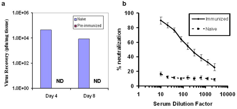
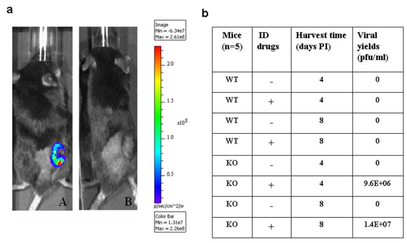
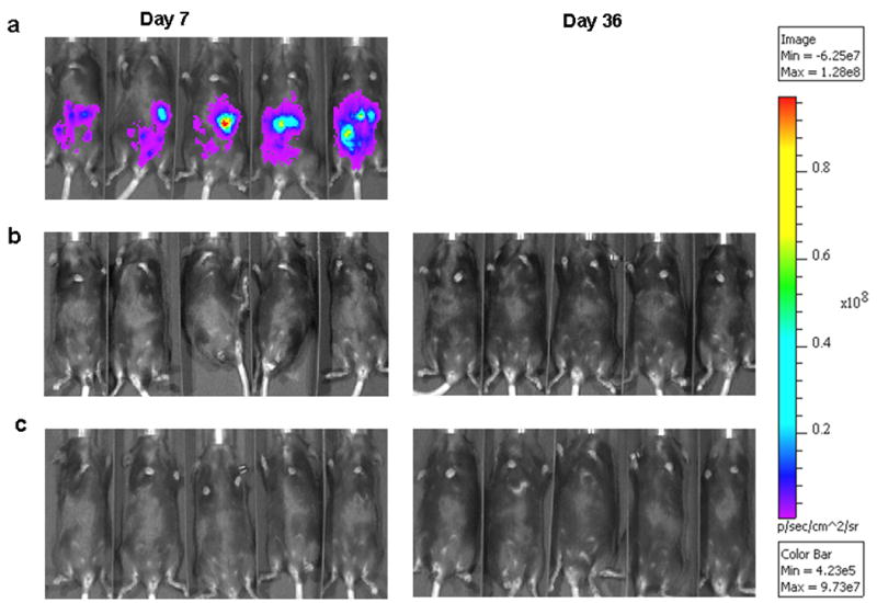
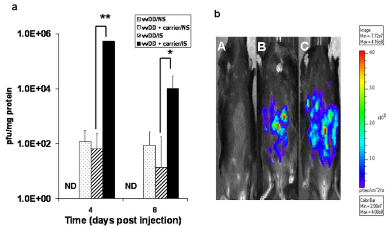
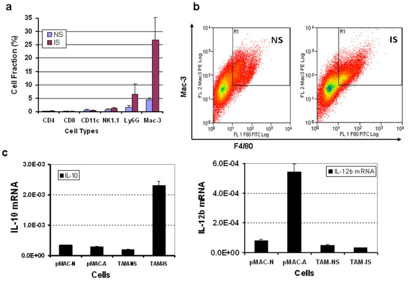
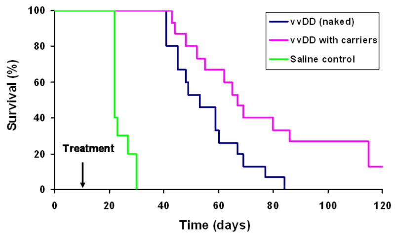
Pre-existing antipoxvirus immunity in cancer patients presents a severe barrier to poxvirus-mediated oncolytic virotherapy. We have explored strategies of immunosuppression (IS) and/or immune evasion for efficient delivery of an oncolytic double-deleted vaccinia virus (vvDD) to tumors in the pre-immunized mice. Transient IS using immunosuppressive drugs, including tacrolimus, mycophenolate mofetil and methylprednisolone sodium succinate, have been used successfully in organ transplantation. This drug cocktail alone did not enhance viral recovery from subcutaneous tumor after systemic viral delivery. Using B-cell knockout mice, we confirmed that the neutralizing antibodies had a significant role in preventing poxvirus infection. Using a MC38 peritoneal carcinomatosis model, we found that the combination of IS and tumor cells as carriers led to the most effective viral delivery, viral replication and viral spread inside the tumor mass. We found that our immunosuppressive drug cocktail facilitated recruitment of tumor-associated macrophages and conversion into an immunosuppressive M2 phenotype (interleukin (IL)-10(hi)/IL-12(low)) in the tumor microenvironment. A combination of IS and carrier cells led to significantly prolonged survival in the tumor model. These results showed the feasibility of treating pre-vaccinated patients with peritoneal carcinomatosis using an oncolytic poxvirus and a combined immune intervention strategy.
Conflict of interest statement
Figures
Similar articles
-
A rationally designed A34R mutant oncolytic poxvirus: improved efficacy in peritoneal carcinomatosis.Mol Ther. 2013 May;21(5):1024-33. doi: 10.1038/mt.2013.27. Epub 2013 Feb 26. Mol Ther. 2013. PMID: 23439499 Free PMC article.
-
A novel therapeutic regimen to eradicate established solid tumors with an effective induction of tumor-specific immunity.Clin Cancer Res. 2012 Dec 15;18(24):6679-89. doi: 10.1158/1078-0432.CCR-12-0979. Epub 2012 Oct 22. Clin Cancer Res. 2012. PMID: 23091113
-
IL-21 arming potentiates the anti-tumor activity of an oncolytic vaccinia virus in monotherapy and combination therapy.J Immunother Cancer. 2021 Jan;9(1):e001647. doi: 10.1136/jitc-2020-001647. J Immunother Cancer. 2021. PMID: 33504576 Free PMC article.
-
Targeted and armed oncolytic poxviruses: a novel multi-mechanistic therapeutic class for cancer.Nat Rev Cancer. 2009 Jan;9(1):64-71. doi: 10.1038/nrc2545. Nat Rev Cancer. 2009. PMID: 19104515 Review.
-
Preclinical and clinical trials of oncolytic vaccinia virus in cancer immunotherapy: a comprehensive review.Cancer Biol Med. 2023 Aug 23;20(9):646-61. doi: 10.20892/j.issn.2095-3941.2023.0202. Cancer Biol Med. 2023. PMID: 37615308 Free PMC article. Review.
Cited by
-
Correlates between host and viral transcriptional program associated with different oncolytic vaccinia virus isolates.Hum Gene Ther Methods. 2012 Oct;23(5):285-96. doi: 10.1089/hgtb.2012.057. Epub 2012 Nov 6. Hum Gene Ther Methods. 2012. PMID: 23131031 Free PMC article.
-
Oncolytic viruses and checkpoint inhibitors: combination therapy in clinical trials.Clin Transl Med. 2018 Nov 14;7(1):35. doi: 10.1186/s40169-018-0214-5. Clin Transl Med. 2018. PMID: 30426287 Free PMC article. Review.
-
Partial Deletion of Glycoprotein B5R Enhances Vaccinia Virus Neutralization Escape while Preserving Oncolytic Function.Mol Ther Oncolytics. 2019 May 21;14:159-171. doi: 10.1016/j.omto.2019.05.003. eCollection 2019 Sep 27. Mol Ther Oncolytics. 2019. PMID: 31236440 Free PMC article.
-
Inhibitors of C5 complement enhance vaccinia virus oncolysis.Cancer Gene Ther. 2013 Jun;20(6):342-50. doi: 10.1038/cgt.2013.26. Epub 2013 May 10. Cancer Gene Ther. 2013. PMID: 23661042 Free PMC article.
-
Curative one-shot systemic virotherapy in murine myeloma.Leukemia. 2012 Aug;26(8):1870-8. doi: 10.1038/leu.2012.70. Epub 2012 Mar 19. Leukemia. 2012. PMID: 22425894 Free PMC article.
References
-
- Parato KA, Senger D, Forsyth PA, Bell JC. Recent progress in the battle between oncolytic viruses and tumours. Nat Rev Cancer. 2005;5:965–976. - PubMed
-
- Kirn DH, Thorne SH. Targeted and armed oncolytic poxviruses: a novel multi-mechanistic therapeutic class for cancer. Nat Rev Cancer. 2009;9:64–71. - PubMed
-
- Zhang Q, Yu YA, Wang E, Chen N, Danner RL, Munson PJ, et al. Eradication of solid human breast tumors in nude mice with an intravenously injected light-emitting oncolytic vaccinia virus. Cancer Res. 2007;67:10038–10046. - PubMed
Publication types
MeSH terms
Substances
Grants and funding
LinkOut - more resources
Full Text Sources
Other Literature Sources
