

Structural basis of low-affinity nickel binding to the nickel-responsive transcription factor NikR from Escherichia coli
- PMID: 20704276
- PMCID: PMC2934763
- DOI: 10.1021/bi100923j
Structural basis of low-affinity nickel binding to the nickel-responsive transcription factor NikR from Escherichia coli
Abstract
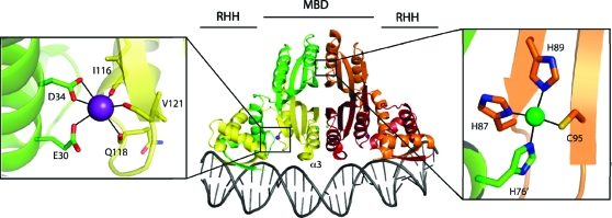
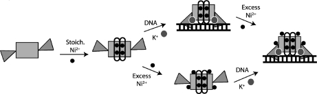
Escherichia coli NikR regulates cellular nickel uptake by binding to the nik operon in the presence of nickel and blocking transcription of genes encoding the nickel uptake transporter. NikR has two binding affinities for the nik operon: a nanomolar dissociation constant with stoichiometric nickel and a picomolar dissociation constant with excess nickel [Bloom, S. L., and Zamble, D. B. (2004) Biochemistry 43, 10029-10038; Chivers, P. T., and Sauer, R. T. (2002) Chem. Biol. 9, 1141-1148]. While it is known that the stoichiometric nickel ions bind at the NikR tetrameric interface [Schreiter, E. R., et al. (2003) Nat. Struct. Biol. 10, 794-799; Schreiter, E. R., et al. (2006) Proc. Natl. Acad. Sci. U.S.A. 103, 13676-13681], the binding sites for excess nickel ions have not been fully described. Here we have determined the crystal structure of NikR in the presence of excess nickel to 2.6 A resolution and have obtained nickel anomalous data (1.4845 A) in the presence of excess nickel for both NikR alone and NikR cocrystallized with a 30-nucleotide piece of double-stranded DNA containing the nik operon. These anomalous data show that excess nickel ions do not bind to a single location on NikR but instead reveal a total of 22 possible low-affinity nickel sites on the NikR tetramer. These sites, for which there are six different types, are all on the surface of NikR, and most are found in both the NikR alone and NikR-DNA structures. Using a combination of crystallographic data and molecular dynamics simulations, the nickel sites can be described as preferring octahedral geometry, utilizing one to three protein ligands (typically histidine) and at least two water molecules.
Figures


Similar articles
-
Metal-selective DNA-binding response of Escherichia coli NikR.Biochemistry. 2004 Aug 10;43(31):10029-38. doi: 10.1021/bi049404k. Biochemistry. 2004. PMID: 15287730
-
Potassium is critical for the Ni(II)-responsive DNA-binding activity of Escherichia coli NikR.J Am Chem Soc. 2010 Feb 10;132(5):1506-7. doi: 10.1021/ja909136h. J Am Chem Soc. 2010. PMID: 20088519
-
Structural basis of the metal specificity for nickel regulatory protein NikR.Biochemistry. 2008 Feb 19;47(7):1938-46. doi: 10.1021/bi702006h. Epub 2008 Jan 15. Biochemistry. 2008. PMID: 18193897 Free PMC article.
-
Coordinating intracellular nickel-metal-site structure-function relationships and the NikR and RcnR repressors.Nat Prod Rep. 2010 May;27(5):658-67. doi: 10.1039/b906683g. Epub 2010 Mar 5. Nat Prod Rep. 2010. PMID: 20442957 Review.
-
Structural determinants of metal selectivity in prokaryotic metal-responsive transcriptional regulators.Biometals. 2005 Aug;18(4):413-28. doi: 10.1007/s10534-005-3716-8. Biometals. 2005. PMID: 16158234 Review.
Cited by
-
Mechanistic insights into the nickel-dependent allosteric response of the Helicobacter pylori NikR transcription factor.J Biol Chem. 2023 Jan;299(1):102785. doi: 10.1016/j.jbc.2022.102785. Epub 2022 Dec 9. J Biol Chem. 2023. PMID: 36502919 Free PMC article.
-
Nickel Metalloregulators and Chaperones.Inorganics (Basel). 2019 Aug;7(8):10.3390/inorganics7080104. doi: 10.3390/inorganics7080104. Epub 2019 Aug 19. Inorganics (Basel). 2019. PMID: 32133383 Free PMC article.
-
Streptomyces coelicolor SCO4226 is a nickel binding protein.PLoS One. 2014 Oct 6;9(10):e109660. doi: 10.1371/journal.pone.0109660. eCollection 2014. PLoS One. 2014. PMID: 25285530 Free PMC article.
-
Metalloprotein Crystallography: More than a Structure.Acc Chem Res. 2016 Apr 19;49(4):695-702. doi: 10.1021/acs.accounts.5b00538. Epub 2016 Mar 15. Acc Chem Res. 2016. PMID: 26975689 Free PMC article.
-
Bacterial Metallostasis: Metal Sensing, Metalloproteome Remodeling, and Metal Trafficking.Chem Rev. 2024 Dec 25;124(24):13574-13659. doi: 10.1021/acs.chemrev.4c00264. Epub 2024 Dec 10. Chem Rev. 2024. PMID: 39658019 Free PMC article. Review.
References
-
- Bloom S. L.; Zamble D. B. (2004) Metal-Selective DNA-Binding Response of Escherichia coli NikR. Biochemistry 43, 10029–10038. - PubMed
-
- Chivers P. T.; Sauer R. T. (2002) NikR Repressor: High-Affinity Nickel Binding to the C-Terminal Domain Regulates Binding to Operator DNA. Chem. Biol. 9, 1141–1148. - PubMed
-
- Schreiter E. R.; Sintchak M. D.; Guo Y.; Chivers P. T.; Sauer R. T.; Drennan C. L. (2003) Crystal structure of nickel-responsive transcription factor NikR. Nat. Struct. Biol. 10, 794–799. - PubMed
Publication types
MeSH terms
Substances
Associated data
- Actions
Grants and funding
LinkOut - more resources
Full Text Sources
Miscellaneous
