. 2010 Aug 12:10:27.
doi: 10.1186/1472-6807-10-27.

Elucidation of the conformational free energy landscape in H.pylori LuxS and its implications to catalysis

Affiliations

Elucidation of the conformational free energy landscape in H.pylori LuxS and its implications to catalysis

Moitrayee Bhattacharyya et al. BMC Struct Biol. .

Abstract

Background: One of the major challenges in understanding enzyme catalysis is to identify the different conformations and their populations at detailed molecular level in response to ligand binding/environment. A detail description of the ligand induced conformational changes provides meaningful insights into the mechanism of action of enzymes and thus its function.

Results: In this study, we have explored the ligand induced conformational changes in H.pylori LuxS and the associated mechanistic features. LuxS, a dimeric protein, produces the precursor (4,5-dihydroxy-2,3-pentanedione) for autoinducer-2 production which is a signalling molecule for bacterial quorum sensing. We have performed molecular dynamics simulations on H.pylori LuxS in its various ligand bound forms and analyzed the simulation trajectories using various techniques including the structure network analysis, free energy evaluation and water dynamics at the active site. The results bring out the mechanistic details such as co-operativity and asymmetry between the two subunits, subtle changes in the conformation as a response to the binding of active and inactive forms of ligands and the population distribution of different conformations in equilibrium. These investigations have enabled us to probe the free energy landscape and identify the corresponding conformations in terms of network parameters. In addition, we have also elucidated the variations in the dynamics of water co-ordination to the Zn2+ ion in LuxS and its relation to the rigidity at the active sites.

Conclusions: In this article, we provide details of a novel method for the identification of conformational changes in the different ligand bound states of the protein, evaluation of ligand-induced free energy changes and the biological relevance of our results in the context of LuxS structure-function. The methodology outlined here is highly generalized to illuminate the linkage between structure and function in any protein of known structure.


Figures

Figure 1
Figure 1
RMSD profiles of LuxS in different ligand bound states. Root-mean-square deviation (RMSD) profiles of LuxS in its various states of ligation [LuxS_apo, LuxS+SRH, LuxS+2SRH, LuxS+KRI, LuxS+2KRI] with reference to the minimized crystal structures.
Figure 2
Figure 2
Dynamically stable hydrogen bonds between protein and ligand. Dynamically stable hydrogen bonds (% of occurrence > 50) for LuxS+2SRH (a) Subunit A and (b) Subunit B and for LuxS+2KRI (c) Subunit A and (d) Subunit B between the protein and ligand. The hydrogen bonds are marked with brown dashed lines. The ligand is depicted in stick representation and the protein residues are represented as slate blue lines. The asymmetry in hydrogen bonding pattern in the two subunits of LuxS+2SRH and LuxS+2KRI around the ribosyl moiety of ligand (SRH/KRI) is highlighted with green rectangular boxes.
Figure 3
Figure 3
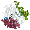
Pictorial representation of the inter subunit correlated and anti-correlated residues of 10 H in LuxS+2KRI. The backbone is represented as cartoon with the two subunits being coloured differently. The correlated and anti-correlated residues are given as maroon and green van der Waals spheres. 10 H is represented as a blue van der Waals sphere. The ligand is given in black sticks representation.
Figure 4
Figure 4
Pictorial representation of the major cliques/communities and water coordination around Zn2+. Pictorial representation of the major cliques and communities in (a) LuxS_apo, (b) LuxS+SRH, (c) LuxS+2SRH, (d) LuxS+KRI, and (e) LuxS+2KRI (upper panels). For LuxS_apo, LuxS+SRH, and LuxS+KRI all communities are plotted whereas for LuxS+2SRH and LuxS+2KRI, only the giant communities around the active sites are plotted for the sake of clarity [upper panel]. The two different subunits are represented as transparent surface and are coloured differently. The clique and community residues are represented as van der Waals spheres and coloured according to the subunit to which they belong. The ligand and Zn2+ are also given in van der Waals representation and coloured red and blue respectively. In the lower panel, water coordination to Zn2+ is depicted for the five systems (LuxS_apo-LuxS+2KRI). The active sites are highlighted in the figure with the Zn2+ being depicted as blue van der Waals spheres and the coordinated water molecules are represented as dark pink sticks. The ligand molecules are depicted as dark grey line representation.
Figure 5
Figure 5
Unique clique residues for the active ligand bound forms of LuxS. The unique clique forming residues in (a) LuxS+KRI w.r.t LuxS+SRH, and (b) LuxS+2KRI w.r.t LuxS+2SRH. The two subunits are given in transparent new cartoon representations and are coloured differently. The unique residues are represented as orange van der Waals spheres and the ones which are near the active sites are depicted as violet van der Waals spheres. It is evident that a larger number of unique clique forming residues are near the active sites of LuxS+2KRI.
Figure 6
Figure 6
Comparison between the hub residues in LuxS_apo-LuxS+2KRI. The central Venn diagram depicts the five systems (LuxS_apo-LuxS+2KRI) as A-E respectively and the different regions are coloured differently. The common regions between LuxS_apo-LuxS+2KRI, LuxS+SRH-LuxS+KRI, and LuxS+2SRH-LuxS+2KRI are coloured deep blue, grey and cyan respectively. The regions which are exclusive to each of LuxS_apo-LuxS+2KRI are highlighted as brown, deep green, yellow, violet, and pink respectively. Three dimensional representations of such comparisons are also given with the two subunits depicted as new cartoon and coloured differently. The common and exclusive hub residues are represented as van der Waals spheres and colour-coded according to the Venn diagram.
Figure 7
Figure 7
Helmholtz free energy contour map and population distribution profile. The Helmholtz free energy contour map for each of the five simulations (on LuxS_apo-LuxS+2KRI, 10 ns each) and the corresponding population distribution in the 'essential plane' are given in inset.
Figure 8
Figure 8
Reversible first step in the reaction between SRH and LuxS. Reversible exchange of the water molecule occupying the fourth coordination site of Zn2+ with the carbonyl group formed upon rearrangement of S-ribosylhomocysteine.

Similar articles

Cited by

References

    1. Monod J, Wyman J, Changeux JP. On the nature of allosteric transitions: a plausible model. J Mol Biol. 1965;12:88–118. doi: 10.1016/S0022-2836(65)80285-6. - DOI - PubMed
    1. Koshland DE, Nemethy G, Filmer D. Comparison of Experimental Binding Data and Theoretical Models in Proteins Containing Subunits. Biochemistry. 1966;5(1):365–385. doi: 10.1021/bi00865a047. - DOI - PubMed
    1. Tsai C, del Sol A, Nussinov R. Allostery: Absence of a change in shape does not imply that allostery is not at play. J Mol Biol. 2008;378:1–11. doi: 10.1016/j.jmb.2008.02.034. - DOI - PMC - PubMed
    1. Tsai C, del Sol A, Nussinov R. Protein allostery, signal transmission and dynamics: a classification scheme of allosteric mechanisms. Mol BioSyst. 2009;5(3):207–216. doi: 10.1039/b819720b. - DOI - PMC - PubMed
    1. Gunasekaran K, Ma B, Nussinov R. Is allostery an intrinsic property of all dynamic proteins? Proteins: Structure, Function, and Bioinformatics. 2004;57:433–443. doi: 10.1002/prot.20232. - DOI - PubMed

Publication types

LinkOut - more resources