Entrapment neuropathy results in different microRNA expression patterns from denervation injury in rats
- PMID: 20704709
- PMCID: PMC2927509
- DOI: 10.1186/1471-2474-11-181
Entrapment neuropathy results in different microRNA expression patterns from denervation injury in rats
Abstract
Background: To compare the microRNA (miRNA) expression profiles in neurons and innervated muscles after sciatic nerve entrapment using a non-constrictive silastic tube, subsequent surgical decompression, and denervation injury.
Methods: The experimental L4-L6 spinal segments, dorsal root ganglia (DRGs), and soleus muscles from each experimental group (sham control, denervation, entrapment, and decompression) were analyzed using an Agilent rat miRNA array to detect dysregulated miRNAs. In addition, muscle-specific miRNAs (miR-1, -133a, and -206) and selectively upregulated miRNAs were subsequently quantified using real-time reverse transcription-polymerase chain reaction (real-time RT-PCR).
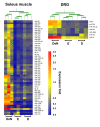
Results: In the soleus muscles, 37 of the 47 miRNAs (13.4% of the 350 unique miRNAs tested) that were significantly downregulated after 6 months of entrapment neuropathy were also among the 40 miRNAs (11.4% of the 350 unique miRNAs tested) that were downregulated after 3 months of decompression. No miRNA was upregulated in both groups. In contrast, only 3 miRNAs were upregulated and 3 miRNAs were downregulated in the denervated muscle after 6 months. In the DRGs, 6 miRNAs in the entrapment group (miR-9, miR-320, miR-324-3p, miR-672, miR-466b, and miR-144) and 3 miRNAs in the decompression group (miR-9, miR-320, and miR-324-3p) were significantly downregulated. No miRNA was upregulated in both groups. We detected 1 downregulated miRNA (miR-144) and 1 upregulated miRNA (miR-21) after sciatic nerve denervation. We were able to separate the muscle or DRG samples into denervation or entrapment neuropathy by performing unsupervised hierarchal clustering analysis. Regarding the muscle-specific miRNAs, real-time RT-PCR analysis revealed an approximately 50% decrease in miR-1 and miR-133a expression levels at 3 and 6 months after entrapment, whereas miR-1 and miR-133a levels were unchanged and were decreased after decompression at 1 and 3 months. In contrast, there were no statistical differences in the expression of miR-206 during nerve entrapment and after decompression. The expression of muscle-specific miRNAs in entrapment neuropathy is different from our previous observations in sciatic nerve denervation injury.
Conclusions: This study revealed the different involvement of miRNAs in neurons and innervated muscles after entrapment neuropathy and denervation injury, and implied that epigenetic regulation is different in these two conditions.
Figures


References
Publication types
MeSH terms
Substances
LinkOut - more resources
Full Text Sources
Other Literature Sources
Medical

