Editorial
. 2010 Aug 13:5:19.
doi: 10.1186/1747-1028-5-19.

Mechanisms of mono- and poly-ubiquitination: Ubiquitination specificity depends on compatibility between the E2 catalytic core and amino acid residues proximal to the lysine

Affiliations
Editorial

Mechanisms of mono- and poly-ubiquitination: Ubiquitination specificity depends on compatibility between the E2 catalytic core and amino acid residues proximal to the lysine

Martin Sadowski et al. Cell Div. .

Abstract

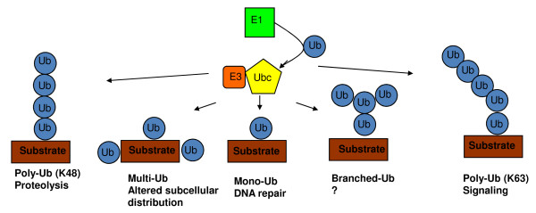
Ubiquitination involves the attachment of ubiquitin to lysine residues on substrate proteins or itself, which can result in protein monoubiquitination or polyubiquitination. Ubiquitin attachment to different lysine residues can generate diverse substrate-ubiquitin structures, targeting proteins to different fates. The mechanisms of lysine selection are not well understood. Ubiquitination by the largest group of E3 ligases, the RING-family E3 s, is catalyzed through co-operation between the non-catalytic ubiquitin-ligase (E3) and the ubiquitin-conjugating enzyme (E2), where the RING E3 binds the substrate and the E2 catalyzes ubiquitin transfer. Previous studies suggest that ubiquitination sites are selected by E3-mediated positioning of the lysine toward the E2 active site. Ultimately, at a catalytic level, ubiquitination of lysine residues within the substrate or ubiquitin occurs by nucleophilic attack of the lysine residue on the thioester bond linking the E2 catalytic cysteine to ubiquitin. One of the best studied RING E3/E2 complexes is the Skp1/Cul1/F box protein complex, SCFCdc4, and its cognate E2, Cdc34, which target the CDK inhibitor Sic1 for K48-linked polyubiquitination, leading to its proteasomal degradation. Our recent studies of this model system demonstrated that residues surrounding Sic1 lysines or lysine 48 in ubiquitin are critical for ubiquitination. This sequence-dependence is linked to evolutionarily conserved key residues in the catalytic region of Cdc34 and can determine if Sic1 is mono- or poly-ubiquitinated. Our studies indicate that amino acid determinants in the Cdc34 catalytic region and their compatibility to those surrounding acceptor lysine residues play important roles in lysine selection. This may represent a general mechanism in directing the mode of ubiquitination in E2 s.


Figures

Figure 1
Figure 1
Different modes of ubiquitination lead to different substrate fates. The versatility of Ub in regulating different processes is derived from its ability to be conjugated as a monomer on one (monoubiquitination) or more substrate lysines (multiubiquitination) or as a polymer (polyubiquitination) by the sequential addition of further Ubs to each other through Ub lysines. Since Ub contains seven lysines, polyubiquitination can generate linear or branched chains with different topologies. Monoubiquitination can regulate DNA repair, viral budding and gene expression, while polyubiquitination through K48 of Ub generally results in proteasomal degradation, and K63-linked Ub chains can function in signaling and endocytosis.
Figure 2
Figure 2
Stereo Images of Ubc9-RanGAP1 Interaction. (A and B) Orthogonal orientations of the Ran-GAP1 consensus motif in complex with Ubc9. Amino acids are indicated by type, number, or both. Ubc9 Glu98 and Asp100 are removed in (B). (C) RanGAP1-Ubc9 interaction outside the motif recognition interface. Helices lettered for RanGAP1 (F and H) and Ubc9 (C). Backbonepositions are represented by ribbon spline (RanGAP1 in yellow, Ubc9 in blue), theoretical hydrogen bonds are dotted lines, and waters are red spheres. *Reprinted from Cell, 108 (3), Bernier-Villamor V, Sampson DA, Matunis MJ and Lima, CD, Structural basis for E2-mediated SUMO conjugation revealed by a complex between ubiquitin-conjugating enzyme Ubc9 and RanGAP1, 345-356., Copyright (2002), with permission from Elsevier.

References

    1. Pickart C, Eddins M. Ubiquitin: structures, functions, mechanisms. Biochim Biophys Acta. 2004;1695:55–72. doi: 10.1016/j.bbamcr.2004.09.019. - DOI - PubMed
    1. Petroski MD, Deshaies RJ. Context of multiubiquitin chain attachment influences the rate of Sic1 degradation. Mol Cell. 2003;11:1435–1444. doi: 10.1016/S1097-2765(03)00221-1. - DOI - PubMed
    1. Petroski M, Deshaies R. Function and regulation of cullin-RING ubiquitin ligases. Nat Rev Mol Cell Biol. 2005;6:8–20. doi: 10.1038/nrm1547. - DOI - PubMed
    1. Passmore LA, Barford D. Getting into position: the catalytic mechanisms of protein ubiquitylation. Biochem J. 2004;379:513–525. doi: 10.1042/BJ20040198. - DOI - PMC - PubMed
    1. Wu G, Xu G, Schulman BA, Jeffrey PD, Harper JW, Pavletich NP. Structure of a beta-TrCP1-Skp1-beta-catenin complex: destruction motif binding and lysine specificity of the SCF(beta-TrCP1) ubiquitin ligase. Mol Cell. 2003;11:1445–1456. doi: 10.1016/S1097-2765(03)00234-X. - DOI - PubMed

Publication types