. 2005 Apr;5(26):213-8.

Quantum dots for molecular imaging and cancer medicine

Affiliations

Quantum dots for molecular imaging and cancer medicine

L A Bentolila et al. Discov Med. 2005 Apr.

Abstract

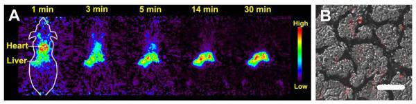
Extract: The past few decades have witnessed technical advances that have introduced cell biologists and physicians to a new, dynamic, subcellular world where genes and gene products can be visualized to interact in space and time and in health and disease. The accelerating field of molecular imaging has been critically dependent on indicator probes which show when and where genetically or biochemically defined molecules, signals or processes appear, interact and disappear, with high spatial and temporal resolution in living cells and whole organisms. For example, the use of radionuclide tracers combined with 3-dimensional (3-D) imaging systems such as Positron Emission Tomography (PET) and Single Photon Emission Computed Tomography (SPECT) are now helping clinicians to characterize the molecular status of tumors deep within patients. Other types of imaging probes rely on the bioluminescence and fluorescence of genetically encoded proteins (originally found in fireflies and jellyfish, respectively) or entirely synthetic fluorochromes, or a combination of both. New powerful biological fluorescence microscopes provide the ability to study single molecules within single cells. Multiphoton confocal microscopy has been developed to allow for the capturing of high-resolution, 3-D images of living tissues that have been tagged with highly specific fluorophores.


Figures

Figure 1
Figure 1
Absorption (upper curves) and emission (lower curves) spectra of four CdSe/ZnS qdot samples. The blue vertical line indicates the emission wavelength of the 488 nm line of an argon ion laser, which can be used to efficiently excite all four types of qdots simultaneously. Inset: a series of CdSe/ZnS dots in aqueous buffer, simultaneously illuminated by a laser (true-color).
Figure 2
Figure 2
Nanocrystal peptide-coating approach. A. Schematic representation of the surface coating chemistry of CdSe/ZnS nanocrystals with phytochelatin-related -peptides. The peptide C-terminal adhesive domain binds to the surface of CdSe/ZnS nanocrystals after exchange with the trioctylphosphine oxide (TOPO) surfactant. A polar and negatively charged hydrophilic linker domain in the peptide sequence provides aqueous buffer solubility to the nanocrystals. TMAOH: Tetramethyl ammonium hydroxide; Cha: 3-cyclohexylalanine. B. Peptide toolkit. The light blue segment contains cysteines and hydrophobic amino acids ensuring binding to the nanocrystal (adhesive domain of Figure 2A) and is common to all peptides. S: solubilization sequence (hydrophilic linker domain of Figure 2A); P: PEG; B: biotin; R: recognition sequence; Q: quencher; D: DOTA (1,4,7,10-tetraazacyclododecane-1,4,7,10-tetraacetic acid) for radionuclide and nuclear spin label chelation; X: any unspecified peptide-encoded function. Qdots solubilization is obtained by a mixture of S and P. Qdots can be targeted with biotin (B), a peptide recognition sequences (R), or other chemical moieties. Qdots fluorescence can be turned on or off by attaching a quencher (Q) via a cleavable peptide link. In the presence of the appropriate enzyme, the quencher is separated from the qdot, restoring the photoluminescence and reporting on the enzyme activity. For simultaneous PET and fluorescence imaging, qdots can be rendered radioactive by chelation of radionuclides using DOTA (D).
Figure 3
Figure 3
MicroPET of 64Cu qdots. Qdots were injected via tail-vein into nude mice and imaged in a small animal scanner. A. Rapid and marked accumulation of qdots in the liver quickly follows their intravenous injection in normal adult nude mice. B. Overlay of a DIC image (Differential Interference Contrast is a popular method of contrast microscopy that utilizes dual-beam interference optics to yield images with a shadowy, three-dimensional relief effect) and a fluorescence image of liver sections showing the accumulation of qdots within cells.
Figure 4
Figure 4
Envisioned future applications for qdots as cancer bioimaging and therapy agents. After injection, qdots homing to the tumor are visualized by PET/MRI/NIR. The tumor is diagnosed in real-time by a confocal catheter or by biopsy and fluorescence analysis. Treatment by specific X-ray or infrared (IR) light absorption by qdots or by free radical photogeneration can follow. Artwork by Lynne C. Olson, UCLA Photographic Services.

References

    1. Alivisatos AP. Semiconductor clusters, nanocrystals, and quantum dots. Science. 1996;271:933–937.
    1. Bruchez M, Moronne M, Gin P, Weiss S, Alivisatos AP. Semiconductor nanocrystals as fluorescent biological labels. Science. 1998;281:2013–2015. - PubMed
    1. Chan WCW, Nie SM. Quantum dot bioconjugates for ultrasensitive nonisotopic detection. Science. 1998;281:2016–2018. - PubMed
    1. Dahan M, Levi S, Luccardini C, Rostaing P, Riveau B, Triller A. Diffusion dynamics of glycine receptors revealed by single-quantum dot tracking. Science. 2003;302:442–445. - PubMed
    1. Kim S, Lim YT, Soltesz EG, De Grand AM, Lee J, Nakayama A, Parker JA, Mihaljevic T, Laurence RG, Dor DM, et al. Near-infrared fluorescent type II quantum dots for sentinel lymph node mapping. Nature Biotechnology. 2004;22:93–97. - PMC - PubMed