

Transcriptional regulation of heart valve development and disease
- PMID: 20705485
- PMCID: PMC2980861
- DOI: 10.1016/j.carpath.2010.06.010
Transcriptional regulation of heart valve development and disease
Abstract
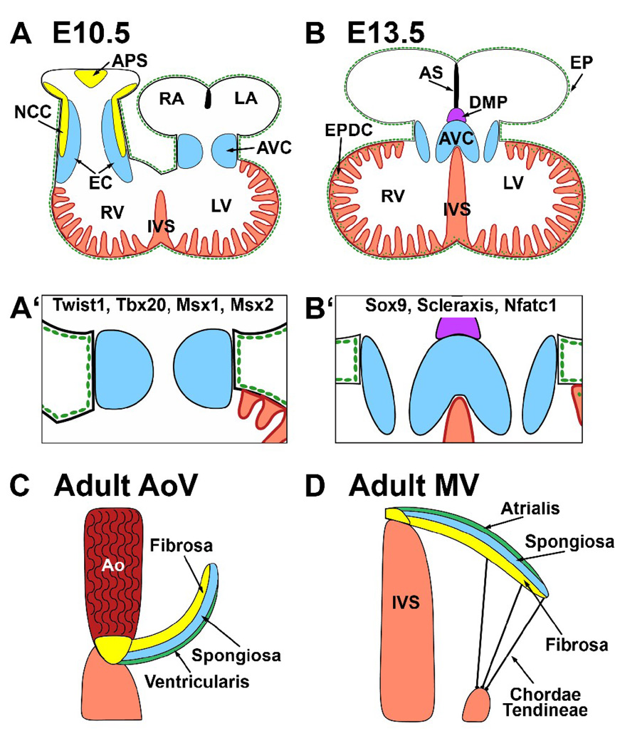
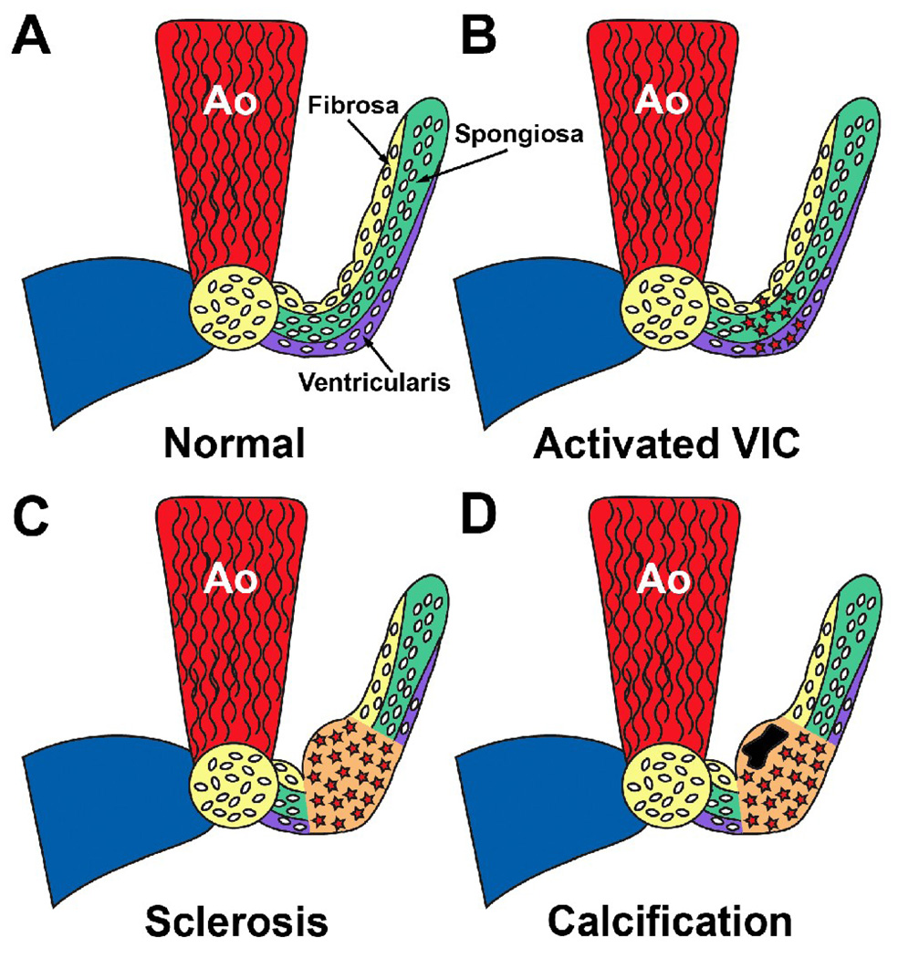
Aortic valve disease is estimated to affect 2% of the United States population. There is increasing evidence that aortic valve disease has a basis in development, as congenital valve malformations are prevalent in patients undergoing valve replacement surgery. In fact, a number of genetic mutations have been linked to valve malformations and disease. In the initial stages of aortic valve pathogenesis, the valvular interstitial cells become activated, undergo cell proliferation, and participate in extracellular matrix remodeling. Many of these cell properties are shared with mesenchymal progenitor cells of the normally developing valves and bones. Historically, valve calcification was thought to be a passive process reflecting end-stage disease. However, recent evidence describes the increased expression of transcription factors in diseased AoV that are common to valvulogenic and osteogenic processes. These studies lend support to the idea that a developmental gene program is reactivated in aortic valve disease and may contribute to the molecular mechanisms underlying valve calcification in disease.
Copyright © 2011 Elsevier Inc. All rights reserved.
Figures
References
-
- Bach DS, Radeva JI, Birnbaum HG, Fournier AA, Tuttle EG. Prevalence, referral patterns, testing, and surgery in aortic valve disease: leaving women and elderly patients behind? J Heart Valve Dis. 2007;16:362–369. - PubMed
-
- Nkomo VT, Gardin JM, Skelton TN, Gottdiener JS, Scott CG, Enriquez-Sarano M. Burden of valvular heart diseases: a population-based study. Lancet. 2006;368:1005–1011. - PubMed
-
- Bonow RO, Carabello BA, Chatterjee K, et al. 2008 Focused update incorporated into the ACC/AHA 2006 guidelines for the management of patients with valvular heart disease: a report of the American College of Cardiology/American Heart Association Task Force on Practice Guidelines (Writing Committee to Revise the 1998 Guidelines for the Management of Patients With Valvular Heart Disease): endorsed by the Society of Cardiovascular Anesthesiologists, Society for Cardiovascular Angiography and Interventions, and Society of Thoracic Surgeons. Circulation. 2008;118:e523–e661. - PubMed
-
- Roberts WC, Ko JM. Frequency by decades of unicuspid, bicuspid, and tricuspid aortic valves in adults having isolated aortic valve replacement for aortic stenosis, with or without associated aortic regurgitation. Circulation. 2005;111:920–925. - PubMed
-
- Lloyd-Jones D, Adams RJ, Brown TM, et al. Heart disease and stroke statistics--2010 update: a report from the American Heart Association. Circulation. 2009;121:e46–e215. - PubMed
Publication types
MeSH terms
Substances
Grants and funding
LinkOut - more resources
Full Text Sources
Medical
