. 2010 Oct 22;285(43):33123-33133.
doi: 10.1074/jbc.M110.139170. Epub 2010 Aug 12.

ABCG2 transports and transfers heme to albumin through its large extracellular loop

Affiliations

ABCG2 transports and transfers heme to albumin through its large extracellular loop

Elodie Desuzinges-Mandon et al. J Biol Chem. .

Abstract

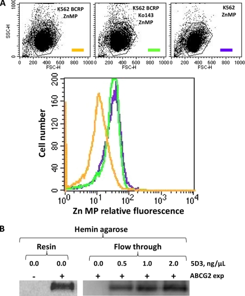
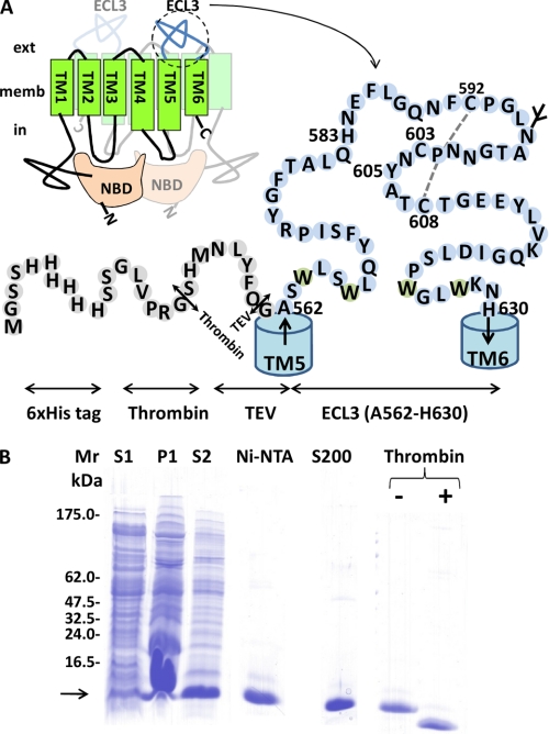
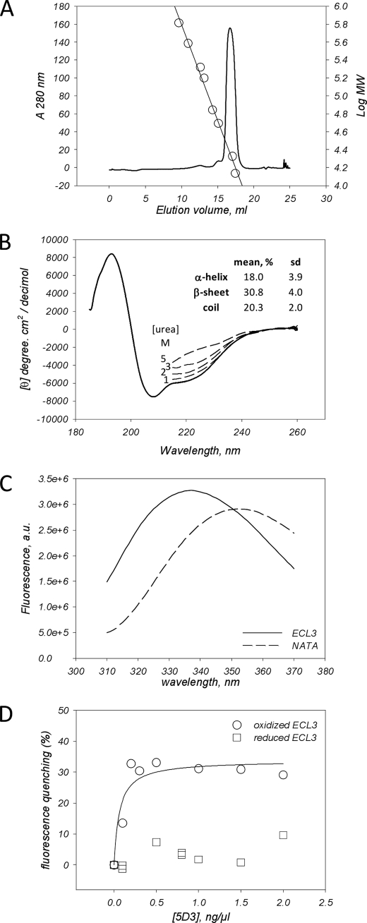
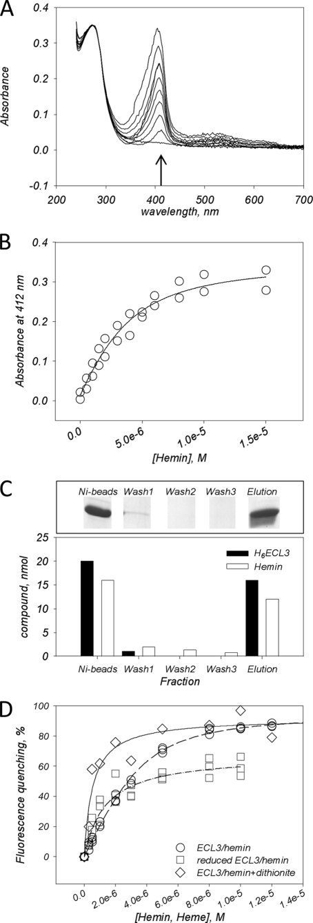
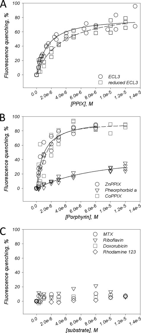
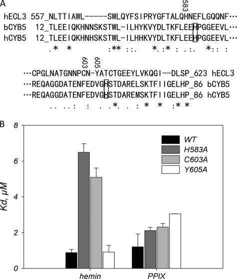
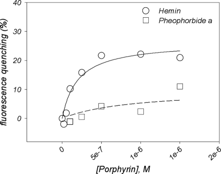
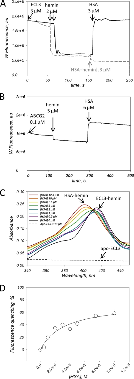
ABCG2 is an ATP-binding cassette (ABC) transporter preferentially expressed by immature human hematopoietic progenitors. Due to its role in drug resistance, its expression has been correlated with a protection role against protoporhyrin IX (PPIX) accumulation in stem cells under hypoxic conditions. We show here that zinc mesoporphyrin, a validated fluorescent heme analog, is transported by ABCG2. We also show that the ABCG2 large extracellular loop ECL3 constitutes a porphyrin-binding domain, which strongly interacts with heme, hemin, PPIX, ZnPPIX, CoPPIX, and much less efficiently with pheophorbide a, but not with vitamin B12. K(d) values are in the range 0.5-3.5 μm, with heme displaying the highest affinity. Nonporphyrin substrates of ABCG2, such as mitoxantrone, doxo/daunorubicin, and riboflavin, do not bind to ECL3. Single-point mutations H583A and C603A inside ECL3 prevent the binding of hemin but hardly affect that of iron-free PPIX. The extracellular location of ECL3 downstream from the transport sites suggests that, after membrane translocation, hemin is transferred to ECL3, which is strategically positioned to release the bound porphyrin to extracellular partners. We show here that human serum albumin could be one of these possible partners as it removes hemin bound to ECL3 and interacts with ABCG2, with a K(d) of about 3 μm.


Figures

FIGURE 1.
FIGURE 1.
Efflux of the heme analog Zn mesoporphyrin (ZnMP) by ABCG2-expressing cells, and decrease of ABCG2 binding to hemin-agarose by the 5D3 mAb raised against ECL3. A, ABCG2-expressing K562 cell transport of ZnMP (orange) compared with K562 control cells (green). Addition of the specific ABCG2 inhibitor Ko143 (1 μm) blocks the efflux of ZnMP (violet). B, modulation by the 5D3 mAb of in vitro ABCG2 binding to hemin-agarose. Whole cell lysates were subjected to a pulldown assay with hemin-agarose in the absence (Resin) or presence of increasing concentrations of 5D3 mAb increasing the amount of unbound ABCG2 (Flow through).
FIGURE 2.
FIGURE 2.
ECL3 design, expression, and purification. A, ECL3 localized between TM spans 5 and 6 extends from Ala562 to His630. It was fused to a His6 N-terminal tag followed by thrombin (Th) and TEV (Te) proteolytic cleavage sites, resulting in a fusion protein of 10,886 Da. The glycosylation site (in mammals) and disulfide bridge between Cys592 and Cys608 are indicated. B, Tricine SDS-PAGE is used to monitor expression and purification of ECL3. S1 and P1 correspond to the supernatant and pellet fractions obtained at low speed centrifugation of bacteria overexpressing the fusion protein. S2 corresponds to the soluble fraction after FC12 solubilization of P1. Ni-NTA and S200 correspond to ECL3 eluted from each resin. The protein was finally digested by thrombin (+ Thrombin) to remove the tag.
FIGURE 3.
FIGURE 3.
ECL3 recombinant domain is folded and recognized by the conformational epitope recognizing 5D3 mAb. A, gel filtration on a Superdex-200 10/300 column of ECL3. The column was calibrated with molecular mass markers (right y log axis, circles) thyroglobulin (650 kDa), ferritin (380 kDa), catalase (210 kDa), aldolase (160 kDa), albumin (70 kDa), ovalbumin (50 kDa), chymotrypsinogen A (21 kDa), and ribonuclease (14 kDa), fitting the elution volume data with SigmaPlot. B, CD spectra of ECL3. The spectrum was fitted for estimating the percentage of each two-dimensional structure (see “Experimental Procedures”). The same experiment was done by adding 1–5 m urea (dashed lines). C, fluorescence spectra of ECL3 (straight line) and NATA (dashed line). D, intrinsic fluorescence quenching of ECL3 (1.5 μm), either oxidized (circles) or reduced (squares), by the 5D3 mAb, as measured at 336 nm and fitted with SigmaPlot using Equation 1.
FIGURE 4.
FIGURE 4.
Hemin and heme binding to ECL3 as monitored by UV absorbance, pulldown assay, and intrinsic fluorescence. A, UV-visible, corrected and normalized, spectra of oxidized hemin (0–15 μm) in the presence of ECL3 (3 μm). B, plot of the absorbance values at 412 nm (arrow in A) of hemin as a function of hemin concentration. C, pulldown assays of the hemin·H6ECL3 complex. The experiment was carried out as described under “Experimental Procedures,” leading to different fractions: Ni-beads, hemin·H6ECL3 complex bound to Ni-NTA magnetic beads; Wash 1–3, magnetic bead washing steps; Elution, imidazole elution step of hemin·H6ECL3 complex. Fractions were loaded on a 16% SDS-PAGE (upper panel), and protein (black bars) and hemin (white bars) amounts were quantified (lower panel). D, intrinsic fluorescence quenching of ECL3 (1–3 μm) with hemin (0–14 μm, circles) and heme as generated by dithionite reduction (0–14 μm, diamonds). Squares correspond to the interaction of hemin with reduced ECL3. Data were fitted using Equation 2 (see “Experimental Procedures”).
FIGURE 5.
FIGURE 5.
Binding of porphyrin and nonporphyrin substrates to ECL3, as monitored by quenching of intrinsic fluorescence. A, binding of PPIX to ECL3, either oxidized (circles) or reduced (squares). B, binding of ZnPPIX (circles), CoPPIX (squares), and pheophorbide a (triangles) to oxidized ECL3. C, Interaction of mitoxantrone (circles), doxorubicin (squares), rhodamine 123 (diamonds), and riboflavin (triangles) with ECL3. Experiments were carried out and analyzed as described in the legend of Fig. 4D.
FIGURE 6.
FIGURE 6.
Single-point mutations H583A and C603A, but not Y605A, in ECL3 dramatically alter hemin binding but hardly affect PPIX binding. A, primary structure alignment of ECL3 with the heme-binding domain of cytochrome b5. The score of homology is as follows: asterisk, identity; colon, strong similarity; period, weak similarity; no symbol for no similarity. Histidine residues of cytochrome b5 involved in iron chelation are boxed. The residues His583, Cys603, and Tyr605, which are replaced by an alanine by site-directed mutagenesis, are numbered. B, dissociation constants for hemin and PPIX binding to H583A, C603A, and Y605A mutant ECL3. Values are taken from the fit of data displayed in supplemental Fig. S3 and summarized in supplemental Table S1.
FIGURE 7.
FIGURE 7.
Effect of previous saturation of ABCG2 drug-binding sites by drug substrates on the ability to bind other substrates further. The full-length transporter was expressed and purified, and fluorescence quenching experiments were carried out as detailed under “Experimental Procedures.” ABCG2 (0.1 μm) was preincubated with 5 μm mitoxantrone and doxorubicin before addition of hemin (circles) or pheophorbide a (squares). Fluorescence experiments were carried out and analyzed as detailed in the legend of Fig. 3C.
FIGURE 8.
FIGURE 8.
Hemin transfer from ECL3 to HSA. A, intrinsic fluorescence of ECL3 (3 μm) was recorded with time before and after addition at the indicated times of 2 μm hemin, and then 3 μm HSA, either alone (black trace) or complexed to hemin (gray dashed trace). ECL3 intrinsic fluorescence was recorded at 336 nm. The contribution of HSA to the fluorescence signal was corrected as detailed in supplemental Fig. S6. B, intrinsic fluorescence of ABCG2 (0.1 μm) preincubated with 5 μm mitoxantrone and doxorubicin was recorded with time before and after addition at the indicated times of 5 μm hemin and then 6 μm HSA. The ABCG2 intrinsic fluorescence was recorded at 328 nm. The contribution of HSA to the fluorescence signal was corrected as detailed in supplemental Fig. S7. C, ECL3 to HSA hemin transfer monitored by UV absorbance. The experiment was carried out as described in Fig. 4 with 10 μm apoECL3 (dashed trace), 5 μm hemin and 0–12.5 μm HSA (black to brown traces). The Soret bands at 402 and 412 nm resulting from the interaction of hemin with HSA and ECL3, respectively, are indicated. D, interaction of HSA with ABCG2 monitored by quenching of ABCG2 intrinsic fluorescence. Fluorescence experiments were carried out and analyzed as detailed in the legend of Fig. 3C, correcting the intrinsic fluorescence of ABCG2 with that of NATA in the presence of the same concentrations of HSA.

Similar articles

Cited by

References

    1. Dean M., Hamon Y., Chimini G. (2001) J. Lipid Res. 42, 1007–1017 - PubMed
    1. Ross D. D., Yang W., Abruzzo L. V., Dalton W. S., Schneider E., Lage H., Dietel M., Greenberger L., Cole S. P., Doyle L. A. (1999) J. Natl. Cancer Inst. 91, 429–433 - PubMed
    1. Litman T., Brangi M., Hudson E., Fetsch P., Abati A., Ross D. D., Miyake K., Resau J. H., Bates S. E. (2000) J. Cell Sci. 113, 2011–2021 - PubMed
    1. Sarkadi B., Ozvegy-Laczka C., Német K., Váradi A. (2004) FEBS Lett. 567, 116–120 - PubMed
    1. Wang X., Baba M. (2005) Antivir. Chem. Chemother. 16, 213–216 - PubMed

Publication types

MeSH terms

LinkOut - more resources