. 2010 Oct 22;285(43):33252-33264.
doi: 10.1074/jbc.M110.146704. Epub 2010 Aug 12.

Effect of trehalose on the properties of mutant {gamma}PKC, which causes spinocerebellar ataxia type 14, in neuronal cell lines and cultured Purkinje cells

Affiliations

Effect of trehalose on the properties of mutant {gamma}PKC, which causes spinocerebellar ataxia type 14, in neuronal cell lines and cultured Purkinje cells

Takahiro Seki et al. J Biol Chem. .

Abstract

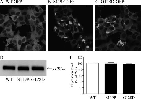
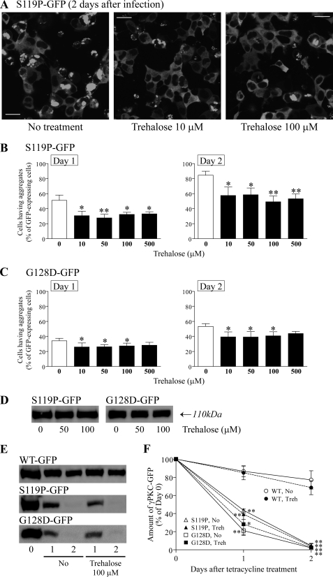
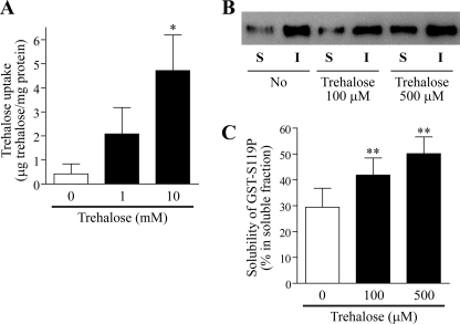
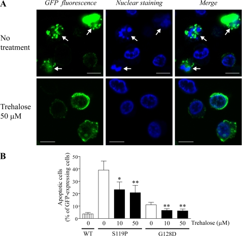
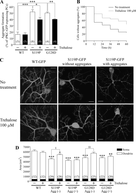
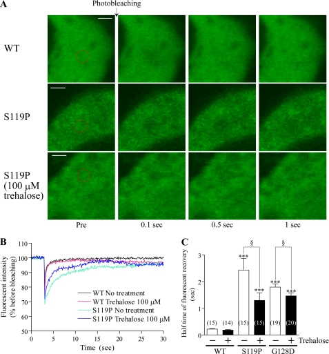
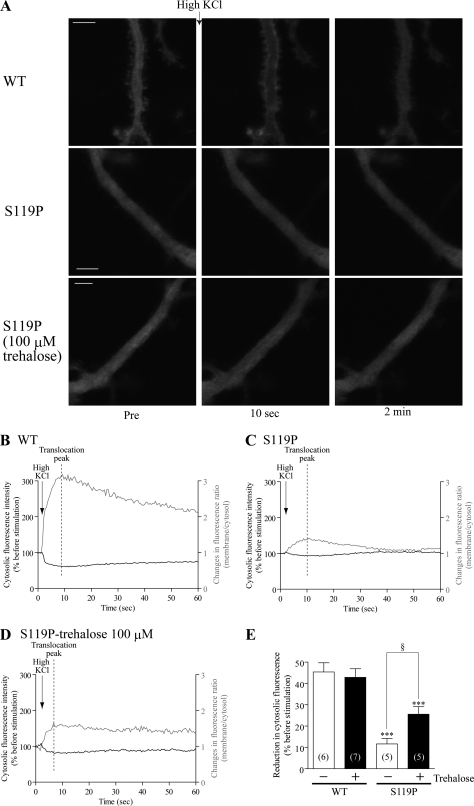
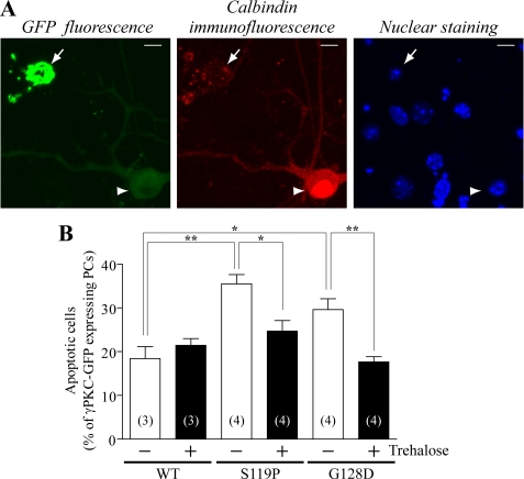
Several missense mutations in the protein kinase Cγ (γPKC) gene have been found to cause spinocerebellar ataxia type 14 (SCA14), an autosomal dominant neurodegenerative disease. We previously demonstrated that the mutant γPKC found in SCA14 is susceptible to aggregation, which induces apoptotic cell death. The disaccharide trehalose has been reported to inhibit aggregate formation and to alleviate symptoms in cellular and animal models of Huntington disease, Alzheimer disease, and prion disease. Here, we show that trehalose can be incorporated into SH-SY5Y cells and reduces the aggregation of mutant γPKC-GFP, thereby inhibiting apoptotic cell death in SH-SY5Y cells and primary cultured Purkinje cells (PCs). Trehalose acts by directly stabilizing the conformation of mutant γPKC without affecting protein turnover. Trehalose was also found to alleviate the improper development of dendrites in PCs expressing mutant γPKC-GFP without aggregates but not in PCs with aggregates. In PCs without aggregates, trehalose improves the mobility and translocation of mutant γPKC-GFP, probably by inhibiting oligomerization and thereby alleviating the improper development of dendrites. These results suggest that trehalose counteracts various cellular dysfunctions that are triggered by mutant γPKC in both neuronal cell lines and primary cultured PCs by inhibiting oligomerization and aggregation of mutant γPKC.


Figures

FIGURE 1.
FIGURE 1.
Expression and aggregation of WT and mutant γPKC-GFP in SH-SY5Y cells infected with adenoviral vectors. A–C, representative images of WT-GFP (A), S119P-GFP (B), and G128D-GFP (C) fluorescence in SH-SY5Y cells at 2 days post-adenoviral infection. No aggregates were found in cells expressing WT-GFP, whereas aggregates of S119P-GFP and G128D-GFP were frequently observed. Bar = 20 μm. D, representative anti-GFP immunoblot of SH-SY5Y cells expressing WT-GFP, S119P-GFP, and G128D-GFP. Cells were harvested 2 days after adenoviral infection. γPKC-GFP was detected with a molecular mass of ∼110 kDa. E, quantitative analysis of immunoblotting in D. Each column represents the mean ± S.E. of three independent experiments. The expression levels of WT-GFP, S119P-GFP, and G128D-GFP were similar in SH-SY5Y cells.
FIGURE 2.
FIGURE 2.
Trehalose inhibits the aggregation of mutant γPKC-GFP in SH-SY5Y cells without affecting its expression level or degradation rate. A, representative images of S119P-GFP fluorescence in SH-SY5Y cells at 2 days post-adenoviral infection without (left) or with trehalose (center: 10 μm, right: 100 μm). Trehalose was added during the adenoviral infection. Trehalose was found to strongly reduce the number of cells with aggregates of S119P-GFP. Bar = 20 μm. B and C, quantitative analyses of the effect of trehalose on aggregation of S119P-GFP (B) and G128D-GFP (C). Left and right graphs indicate the results at 1 and 2 days after adenoviral infection, respectively. We evaluated 100–200 GFP-positive cells in each experiment. Each column represents the mean ± S.E. of the percentages of cells with aggregates. Various concentrations of trehalose significantly inhibit the aggregation of mutant γPKC-GFP in SH-SY5Y cells (*, p < 0.05; **, p < 0.01 versus without trehalose, Dunnett's multiple comparison test, n = 5 in S119P-GFP, n = 4 in G128D-GFP). D, representative anti-GFP immunoblot of SH-SY5Y cells expressing S119P-GFP (left) and G128D-GFP (right). Cells were cultured for 2 days after adenoviral infection without or with trehalose (50 or 100 μm). Trehalose does not affect the expression levels of S119P-GFP and G128D-GFP. E, representative anti-GFP immunoblot results of a chase assay for WT-GFP (upper), S119P-GFP (middle), and G128D-GFP (lower). One day after adenoviral infection, the expression of γPKC-GFP was arrested with the addition of tetracycline (Tet, 1 μg/ml). Trehalose (100 μm) was added at the time of Tet treatment. The number of elapsed days after Tet treatment is indicated below the panels. F, quantitative analyses of the chase assay in E. Each symbol represents the mean ± S.E. of the residual amount of WT-GFP (circle), S119P-GFP (triangle), or G128D-GFP (square), indicated as the percentage of day 0. Open and closed symbols indicate the results of cells without (No) and with 100 μm trehalose (Treh), respectively. S119P-GFP and G128D-GFP were found to be more rapidly degraded than WT-GFP after expression arrest, but trehalose does not significantly affect the degradation rates of WT-GFP, S119P-GFP, or G128D-GFP (*, p < 0.05; **, p < 0.01, unpaired t test, n = 3).
FIGURE 3.
FIGURE 3.
Trehalose is incorporated into SH-SY5Y cells and inhibits transition of recombinant mutant γPKC into the insoluble fraction. A, quantitative analysis of trehalose uptake in SH-SY5Y cells. Cells were cultured for 48 h in the presence of 0–10 mm trehalose, as indicated below the graph. The amount of intracellular trehalose was quantified as described under “Experimental Procedures.” The levels of intracellular trehalose were significantly higher in SH-SY5Y cells cultured with 10 mm trehalose for 48 h (*, p < 0.05 versus without trehalose, Dunnett's multiple comparison test, n = 5). B, representative anti-γPKC-immunoblot results of soluble (S) and insoluble (I) fractions of recombinant GST-S119P after a 1-h incubation at 37 °C without or with trehalose (100 or 500 μm). C, quantitative analysis of immunoblot results after a 1-h incubation in B. Each column represents the mean ± S.E. of the percentage of GST-S119P found in S fraction relative to the total amount of GST-S119P (S + I fraction). Trehalose significantly inhibited insolubilization of recombinant GST-S119P by a 1-h incubation at 37 °C (**, p < 0.01, Dunnett's multiple comparison test, n = 4).
FIGURE 4.
FIGURE 4.
Trehalose alleviates cytotoxicity induced by mutant γPKC-GFP in SH-SY5Y cells. Effect of trehalose on apoptosis induced by mutant γPKC. Three days after adenoviral infection, SH-SY5Y cells were stained with Hoechst 33342 (50 μg/ml). Apoptosis was evaluated by examining chromatin condensation and fragmentation of the nuclei stained by Hoechst 33342. A, representative images of GFP (left), Hoechst 33342 (center), and merged fluorescence of cells expressing S119P-GFP in the absence (upper) or presence (lower) of 50 μm trehalose. Arrows in the upper images indicate apoptotic cells with condensed or fragmented nuclei. Bar = 10 μm. B, quantitative analyses of the apoptotic cells by nuclear staining in B. Each column represents the mean ± S.E. of the percentage of apoptotic cells in the GFP-positive population. Trehalose (10 and 50 μm) significantly reduces the number of apoptotic cells expressing S119P-GFP and G128D-GFP (*, p < 0.05; **, p < 0.01 versus without trehalose, Dunnett's multiple comparison test, n = 4).
FIGURE 5.
FIGURE 5.
Trehalose inhibits aggregation of mutant γPKC-GFP and mutant γPKC-GFP-mediated incorrect development of dendrites in primary cultured PCs. A, effect of trehalose on aggregation of WT-GFP, S119P-GFP, and G128D-GFP in primary cultured PCs. Adenoviral vectors were infected on DIV14, at the same time of trehalose (100 μm) treatment, and GFP fluorescence was observed on DIV28. Each column represents the mean ± S.E. of the percentage of cells with aggregates of γPKC-GFP in GFP-positive PCs. The numbers of experiments are indicated in the columns. S119P-GFP and G128D-GFP forms aggregates in PCs more frequently than WT-GFP, and trehalose significantly inhibits of the aggregation of S119P-GFP and G128D-GFP in PCs (*, p < 0.05; ***, p < 0.001, unpaired t test). B, effect of trehalose on aggregation of S119P-GFP in PCs evaluated by long term time-lapse imaging. Adenoviral vectors were infected on DIV22, at the same time of trehalose (100 μm) treatment, and the GFP fluorescence was acquired every 30 min for 60 h from DIV26 to DIV29. Aggregation of S119P-GFP was evaluated every 6 h. The black line (no treatment, 73 cells) and gray line (trehalose 100 μm, 33 cells) indicate the percentages of PCs that never formed aggregates during the observation period. Trehalose significantly delays aggregate formation of S119P-GFP in PCs (p < 0.01, log rank test). C and D, effect of trehalose on dendrite morphology in PCs expressing WT-GFP, S119P-GFP, and G128D-GFP. Adenoviral vectors were infected on DIV14, at the same time of trehalose (100 μm) treatment, and GFP fluorescence was observed on DIV28. C, representative images of GFP fluorescence in PCs expressing WT-GFP (left), S119P-GFP without aggregates (center), and S119P-GFP with aggregates (right). Upper and lower images show PCs without and with 100 μm trehalose treatment. D, quantitative analysis of areas of the PCs expressing WT-GFP, S119P-GFP, and G128D-GFP. Each column represents the mean ± S.E. of the PC areas, which were quantified by measuring the distribution of GFP fluorescence. The numbers of observed PCs are indicated in the columns. Open and closed portions of the columns indicate the areas of the PC dendrites and somata, respectively. Trehalose significantly prevents the decrease in area, especially in the dendrites, of PCs expressing S119P-GFP and G128D-GFP without aggregates; the areas of PCs with S119P-GFP and G128D-GFP aggregates were not affected by trehalose (*, p < 0.05; **, p < 0.01; ***, p < 0.001 versus WT-GFP with no treatment, §, p < 0.05; §§, p < 0.01, unpaired t test).
FIGURE 6.
FIGURE 6.
Trehalose increases the mobility of mutant γPKC-GFP in primary cultured PCs. A, representative images of GFP fluorescence in PC somata expressing WT-GFP (upper panels), S119P-GFP (middle panels), or S119P-GFP cultured with 100 μm trehalose (lower panels) before (Pre) and 0.1, 0.5, and 2 s after photobleaching. Bleached areas are shown as red circles. Cells were infected on DIV14 and observed on DIV28. Bar = 2 μm. B, temporal changes in fluorescence intensities in bleached areas of PC somata expressing WT-GFP and S119P-GFP with or without trehalose. C, half time of fluorescence recovery in bleached area of PC somata expressing WT-GFP, S119P-GFP, and G128D-GFP with or without trehalose. Half time of recovery was calculated by fitting the changes in fluorescence intensities after photobleaching to single exponential functions. Data represent means ± S.E. of half times obtained from 14–20 PCs. Trehalose significantly decreases the half times of S119P-GFP and G128D-GFP in PC somata (***, p < 0.001 versus WT; §, p < 0.05, unpaired t test).
FIGURE 7.
FIGURE 7.
Trehalose prevents attenuated translocation of mutant γPKC-GFP in primary cultured PCs. A, representative images of GFP fluorescence in PC dendrites expressing WT-GFP (upper panels) and S119P-GFP (middle panels) and S119P-GFP cultured with 100 μm trehalose (lower panels) before (Pre) or 10 s and 2 min after high KCl (100 mm KCl, 50 μl) stimulation. PCs were infected on DIV14 and observed on DIV28. Bar = 5 μm. B–D, temporal changes in fluorescent intensity (black line) and fluorescent ratio (membrane/cytosol, gray line) of WT-GFP (B), S119-GFP (C), and S119P-GFP in the presence of 100 μm trehalose (D) in PC dendrites shown in A. We evaluated the reduction in fluorescence intensity of cytosolic γPKC-GFP at the translocation peak, the time point when the fluorescence ratio reached maximum, as the index for translocation amplitude (B–D). E, translocation amplitude was quantitatively evaluated by measuring the reduction in fluorescence of cytosolic γPKC-GFP. Data represent means ± S.E. of amplitudes obtained from five to seven PCs. Trehalose significantly improves the translocation amplitude of S119P-GFP in PCs (***, p < 0.001 versus WT; §, p < 0.05, unpaired t test).
FIGURE 8.
FIGURE 8.
Trehalose inhibits apoptotic cell death in PCs expressing a mutant γPKC-GFP. A, representative GFP fluorescence (left), calbindin immunostaining (center), and nuclear staining (right) in PCs expressing S119P-GFP. Cells were infected on DIV14, fixed on DIV28, and immunostained with an anti-calbindin antibody, concomitantly with nuclear staining with Hoechst 33342 (0.5 μg/ml). Cells with fragmented or condensed nuclei were regarded as apoptotic cells (arrow). All apoptotic PCs had aggregates of mutant γPKC-GFP and stained with the anti-calbindin antibody in a dot-like manner. The arrowhead indicates a surviving PC lacking aggregates of S119P-GFP and containing a normal nucleus. Bar = 10 μm. B, quantitative analysis of apoptotic cells by nuclear staining in A. Each column represents the mean ± S.E. of the percentage of apoptotic cells in GFP-positive PCs. The number of experiments is indicated in each column. S119P-GFP and G128D-GFP triggers apoptosis in PCs, but trehalose (100 μm) significantly reduces the number of apoptotic cells in the population expressing mutant γPKC-GFP (*, p < 0.05; **, p < 0.01, unpaired t test).

Similar articles

Cited by

References

    1. Schöls L., Bauer P., Schmidt T., Schulte T., Riess O. (2004) Lancet Neurol. 3, 291–304 - PubMed
    1. Cagnoli C., Mariotti C., Taroni F., Seri M., Brussino A., Michielotto C., Grisoli M., Di Bella D., Migone N., Gellera C., Di Donato S., Brusco A. (2006) Brain 129, 235–242 - PubMed
    1. Dueñas A. M., Goold R., Giunti P. (2006) Brain 129, 1357–1370 - PubMed
    1. Martin J. B. (1999) N. Engl. J. Med. 340, 1970–1980 - PubMed
    1. Everett C. M., Wood N. W. (2004) Brain 127, 2385–2405 - PubMed

Publication types

MeSH terms

LinkOut - more resources