

Interfacial kinetic and binding properties of mammalian group IVB phospholipase A2 (cPLA2beta) and comparison with the other cPLA2 isoforms
- PMID: 20705608
- PMCID: PMC2975232
- DOI: 10.1074/jbc.M110.165647
Interfacial kinetic and binding properties of mammalian group IVB phospholipase A2 (cPLA2beta) and comparison with the other cPLA2 isoforms
Abstract
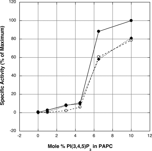
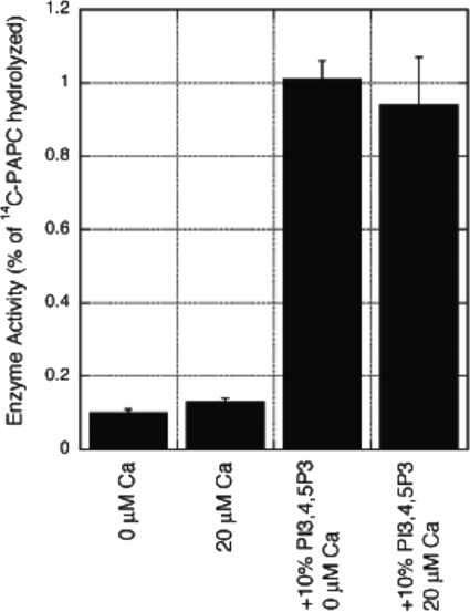
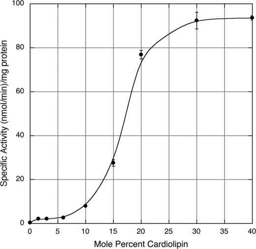
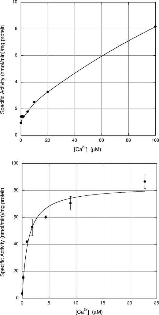
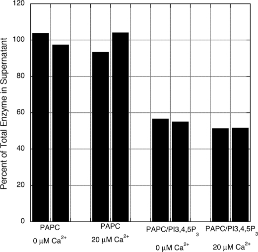
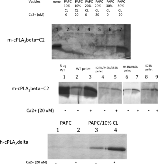
The cytosolic (group IV) phospholipase A(2) (cPLA(2)s) family contains six members. We have prepared recombinant proteins for human α, mouse β, human γ, human δ, human ε, and mouse ζ cPLA(2)s and have studied their interfacial kinetic and binding properties in vitro. Mouse cPLA(2)β action on phosphatidylcholine vesicles is activated by anionic phosphoinositides and cardiolipin but displays a requirement for Ca(2+) only in the presence of cardiolipin. This activation pattern is explained by the effects of anionic phospholipids and Ca(2+) on the interfacial binding of mouse cPLA(2)β and its C2 domain to vesicles. Ca(2+)-dependent binding of mouse cPLA(2)β to cardiolipin-containing vesicles requires a patch of basic residues near the Ca(2+)-binding surface loops of the C2 domain, but binding to phosphoinositide-containing vesicles does not depend on any specific cluster of basic residues. Human cPLA(2)δ also displays Ca(2+)- and cardiolipin-enhanced interfacial binding and activity. The lysophospholipase, phospholipase A(1), and phospholipase A(2) activities of the full set of mammalian cPLA(2)s were quantified. The relative level of these activities is very different among the isoforms, and human cPLA(2)δ stands out as having relatively high phospholipase A(1) activity. We also tested the susceptibility of all cPLA(2) family members to a panel of previously reported inhibitors of human cPLA(2)α and analogs of these compounds. This led to the discovery of a potent and selective inhibitor of mouse cPLA(2)β. These in vitro studies help determine the regulation and function of the cPLA(2) family members.
Figures
Similar articles
-
C2-di-ethyl-ceramide-1-phosphate as an inhibitor of group IVA cytosolic phospholipase A2.Eur J Pharmacol. 2012 Dec 15;697(1-3):144-51. doi: 10.1016/j.ejphar.2012.09.041. Epub 2012 Oct 5. Eur J Pharmacol. 2012. PMID: 23043861
-
Identification of novel cytosolic phospholipase A(2)s, murine cPLA(2){delta}, {epsilon}, and {zeta}, which form a gene cluster with cPLA(2){beta}.J Biol Chem. 2005 Jul 1;280(26):24576-83. doi: 10.1074/jbc.M413711200. Epub 2005 May 2. J Biol Chem. 2005. PMID: 15866882
-
Role of phosphorylation and basic residues in the catalytic domain of cytosolic phospholipase A2alpha in regulating interfacial kinetics and binding and cellular function.J Biol Chem. 2009 Apr 3;284(14):9596-611. doi: 10.1074/jbc.M807299200. Epub 2009 Jan 28. J Biol Chem. 2009. PMID: 19176526 Free PMC article.
-
Localization and function of cytosolic phospholipase A2alpha at the Golgi.Biochimie. 2010 Jun;92(6):620-6. doi: 10.1016/j.biochi.2010.03.001. Epub 2010 Mar 10. Biochimie. 2010. PMID: 20226226 Free PMC article. Review.
-
Involvement of cytosolic phospholipase A(2), calcium independent phospholipase A(2) and plasmalogen selective phospholipase A(2) in neurodegenerative and neuropsychiatric conditions.Curr Med Chem. 2010;17(25):2746-63. doi: 10.2174/092986710791859289. Curr Med Chem. 2010. PMID: 20586719 Review.
Cited by
-
Function of SYDE C2-RhoGAP family as signaling hubs for neuronal development deduced by computational analysis.Sci Rep. 2022 Mar 12;12(1):4325. doi: 10.1038/s41598-022-08147-7. Sci Rep. 2022. PMID: 35279680 Free PMC article.
-
Phospholipase A2 enzymes: physical structure, biological function, disease implication, chemical inhibition, and therapeutic intervention.Chem Rev. 2011 Oct 12;111(10):6130-85. doi: 10.1021/cr200085w. Epub 2011 Sep 12. Chem Rev. 2011. PMID: 21910409 Free PMC article. Review. No abstract available.
-
Lipid-metabolizing serine hydrolases in the mammalian central nervous system: endocannabinoids and beyond.Biochim Biophys Acta Mol Cell Biol Lipids. 2019 Jun;1864(6):907-921. doi: 10.1016/j.bbalip.2018.08.007. Epub 2018 Aug 16. Biochim Biophys Acta Mol Cell Biol Lipids. 2019. PMID: 30905349 Free PMC article. Review.
-
A calcium-dependent acyltransferase that produces N-acyl phosphatidylethanolamines.Nat Chem Biol. 2016 Sep;12(9):669-71. doi: 10.1038/nchembio.2127. Epub 2016 Jul 11. Nat Chem Biol. 2016. PMID: 27399000 Free PMC article.
-
Reduced kidney levels of lysophosphatidic acids in rats after chronic administration of aristolochic acid: Its possible protective role in renal fibrosis.Toxicol Rep. 2015 Feb 28;2:121-129. doi: 10.1016/j.toxrep.2015.02.012. eCollection 2015. Toxicol Rep. 2015. PMID: 28962344 Free PMC article.
References
-
- Ohto T., Uozumi N., Hirabayashi T., Shimizu T. (2005) J. Biol. Chem. 280, 24576–24583 - PubMed
-
- Ghosh M., Tucker D. E., Burchett S. A., Leslie C. C. (2006) Prog. Lipid Res. 45, 487–510 - PubMed
-
- Ghosh M., Loper R., Gelb M. H., Leslie C. C. (2006) J. Biol. Chem. 281, 16615–16624 - PubMed
-
- de Carvalho M. G., Garritano J., Leslie C. C. (1995) J. Biol. Chem. 270, 20439–20446 - PubMed
Publication types
MeSH terms
Substances
Grants and funding
LinkOut - more resources
Full Text Sources
Molecular Biology Databases
Miscellaneous
