Review
. 2011 Mar-Apr;17(2):254-71.
doi: 10.1093/humupd/dmq035. Epub 2010 Aug 12.

The different shades of mammalian pluripotent stem cells

Affiliations
Review

The different shades of mammalian pluripotent stem cells

Ewart W Kuijk et al. Hum Reprod Update. 2011 Mar-Apr.

Abstract

Background: Pluripotent stem cells have been derived from a variety of sources such as from the inner cell mass of preimplantation embryos, from primordial germ cells, from teratocarcinomas and from male germ cells. The recent development of induced pluripotent stem cells demonstrates that somatic cells can be reprogrammed to a pluripotent state in vitro.

Methods: This review summarizes our current understanding of the origins of mouse and human pluripotent cells. We pay specific attention to transcriptional and epigenetic regulation in pluripotent cells and germ cells. Furthermore, we discuss developmental aspects in the germline that seem to be of importance for the transition of germ cells towards pluripotency. This review is based on literature from the Pubmed database, using Boolean search statements with relevant keywords on the subject.

Results: There are distinct molecular mechanisms involved in the generation and maintenance of the various pluripotent cell types. Furthermore, there are important similarities and differences between the different categories of pluripotent cells in terms of phenotype and epigenetic modifications. Pluripotent cell lines from various origins differ in growth characteristics, developmental potential, transcriptional activity and epigenetic regulation. Upon derivation, pluripotent stem cells generally acquire new properties, but they often also retain a 'footprint' of their tissue of origin.

Conclusions: In order to further our knowledge of the mechanisms underlying self-renewal and pluripotency, a thorough comparison between different pluripotent stem cell types is required. This will progress the use of stem cells in basic biology, drug discovery and future clinical applications.


Figures

Figure 1
Figure 1
Schematic representation of the cell signal transduction pathways that are involved in pluripotency of mouse and human ES cells. Mouse ES cells: LIF binds to LIFR and GP130 heterodimers, which results in the activation of STAT3 signalling and AKT signalling and subsequent activation of the downstream target genes Klf4 and Tbx3. BMP binds to heterodimers of the Type I and Type II receptors. As a result, three Smad transcription factors (one of which is Smad 4) trimerize and translocate to the nucleus where they activate the expression of Id genes. LIF and BMP signalling also results in the expression of other members of the pluripotency factor network (e.g. Sox2, Nanog and Pou5f1), but it is unclear if this is a direct or an indirect effect. Human ES cells: FGF binds to FGF-receptor homodimers, which leads to AKT signalling. In parallel, Activin or Nodal homodimers binds to heterodimers of the Type I and Type II receptors. As a result, three Smad transcription factors (one of which is Smad 4) trimerize and translocate to the nucleus. Cooperatively, FGF and Activin/Nodal maintain pluripotency of human ES cells. The question marks depict our lack of knowledge on how these cell signal transduction pathways act on the pluripotency factor network (dashed line) in human ES cells.
Figure 2
Figure 2
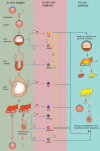
Origins of mammalian pluripotent cells. In the green column on the left are the developmental stages and in vivo origins from which mammalian pluripotent stem cells have been derived. In the pink column in the middle are the stem cell types that can be propagated in vitro. In the blue column on the right is indicated which in vitro cultured mouse cells can participate in all three germ layers including germ cells as determined by the chimera assay. In the same column is indicated which in vitro cultured mouse cell types can restore spermatogenesis as determined by the testis transplantation assay. Blue arrows indicate the derivation of in vitro cell lines from in vivo origins, yellow arrows indicate in vitro transformation/differentiation of one cell type into another, green arrows indicate in vivo functionality tests of in vitro cultured cells. White numbered squares refer to studies performed in mice and red numbered squares refer to studies performed in human: 1Evans and Kaufman (1981), Martin (1981); 2Thomson et al. (1998); 3Chou et al. (2008); 4Brons et al. (2007), Tesar et al. (2007); 5Matsui et al. (1992), Resnick et al. (1992); 6Liu et al. (2004), Shamblott et al. (1998), Turnpenny et al. (2003); 7Okita et al. (2007), Takahashi and Yamanaka (2006); 8Takahashi et al. (2007); 9Kanatsu-Shinohara et al. (2004), Ko et al. (2009), Seandel et al. (2007); 10Conrad et al. (2008), Golestaneh et al. (2009), Kossack et al. (2009), Mizrak et al. (2009); 11Guan et al. (2006); 12Kanatsu-Shinohara et al. (2003); 13Bao et al. (2009), Guo et al. (2009), Silva et al. (2009); 14Guo et al. (2009); 15Nayernia et al. (2006).
Figure 3
Figure 3
The first two lineage segregation events in mammals. From left to right: three successive stages (morula, early blastocyst and late blastocyst) in mammalian development. The two consecutive lineage segregation events that occur in this developmental period are schematically depicted below these embryonic stages. The first segregation of cell lineages occurs in morula stage embryos and results in the formation of the trophectoderm (brown) and the cells of the ICM (red). In the ICM, this event is followed by a second segregation event that initially results in the formation of randomly distributed primitive endoderm precursors (green) and epiblast precursors (red). These cells are subsequently sorted out to form the primitive endoderm and the pluripotent epiblast at the late blastocyst stage.
Figure 4
Figure 4
The segregation of the trophectoderm from the ICM. The segregation of the trophectoderm and the ICM is governed by Hippo signalling. In the outer cells of the embryo (left). Hippo signalling is suppressed possibly as a result of a signal generated by cell polarity. YAP is translocated to the nucleas where it co-operates with TEAD4 to induce the expression of the trophectoderm genes Cdx2 and Gata3, which in turn leads to the induction of a trophoblast gene expression program and the down-regulation of POU5F1. In inner cells (right), Hippo signalling is active resulting in LATS-mediated phosphorylation of YAP. Phosphorylated YAP is sequestered in the cytoplasm. In the absence of nuclear YAP localization, TEAD4 remains inactive, the trophectoderm genes Cdx2 and Gata3 are consequently not expressed, and initial POU5F1 expression is maintained (Nishioka et al., 2009).
Figure 5
Figure 5
The segregation of the primitive endoderm from the epiblast. Schematic representation of the ICM of E3.5 (top-right) and E4.5 embryos (bottom-right). In E3.5 embryos, cellular differences in FGF-signalling (enlarged cells on the left) result in a heterogeneous ICM with NANOG-positive (red) and GATA6-positive (green) cells, which are the precursors for the epiblast (EPI) and the primitive endoderm (PE) respectively. Extracellular matrix (ECM) genes are up-regulated and the cells are sorted out to form the primitive endoderm and epiblast lineages at E4.5 (Chazaud et al., 2006).
Figure 6
Figure 6
A network controlling pluripotency. The transcription factors POU5F1, SOX2 and NANOG bind to the same promoter regions of a large number of genes, thereby regulating transcriptional activity of these genes (Boyer et al., 2005; Loh et al., 2006; Chen et al., 2008; Sharov et al., 2008). The cooperative binding of these factors results in the activation of genes that code for transcription factors, signal transduction components, and epigenetic modifiers that promote pluripotency and self-renewal, including Pou5f1, Sox2 and Nanog (arrows). In addition, POU5F1, SOX2 and NANOG bind to the promotors of genes that are silent in ES cells, expression of which is associated with lineage commitment and differentiation (blunted arrows).

Similar articles

Cited by

References

    1. Andrews PW. Retinoic acid induces neuronal differentiation of a cloned human embryonal carcinoma cell line in vitro. Dev Biol. 1984;103:285–293. doi:10.1016/0012-1606(84)90316-6. - DOI - PubMed
    1. Andrews PW. Human teratocarcinomas. Biochim Biophys Acta. 1988;948:17–36. - PubMed
    1. Andrews PW. From teratocarcinomas to embryonic stem cells. Philos Trans R Soc Lond B Biol Sci. 2002;357:405–417. - PMC - PubMed
    1. Andrews PW, Damjanov I, Simon D, Banting GS, Carlin C, Dracopoli NC, Fogh J. Pluripotent embryonal carcinoma clones derived from the human teratocarcinoma cell line Tera-2. Differentiation in vivo and in vitro. Lab Invest. 1984;50:147–162. - PubMed
    1. Arman E, Haffner-Krausz R, Chen Y, Heath JK, Lonai P. Targeted disruption of fibroblast growth factor (FGF) receptor 2 suggests a role for FGF signaling in pregastrulation mammalian development. Proc Natl Acad Sci USA. 1998;95:5082–5087. doi:10.1073/pnas.95.9.5082. - DOI - PMC - PubMed

Publication types