. 2010 Nov 18;116(20):4317-27.
doi: 10.1182/blood-2010-01-261206. Epub 2010 Aug 12.

Platelet CD40L mediates thrombotic and inflammatory processes in atherosclerosis

Affiliations

Platelet CD40L mediates thrombotic and inflammatory processes in atherosclerosis

Dirk Lievens et al. Blood. .

Abstract

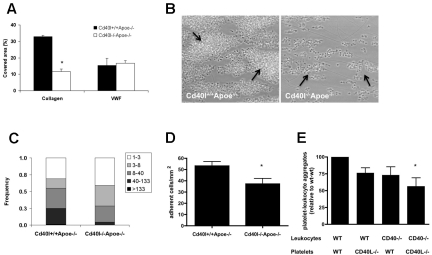
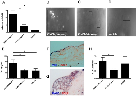
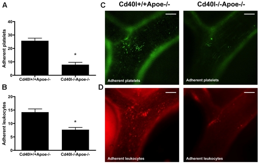
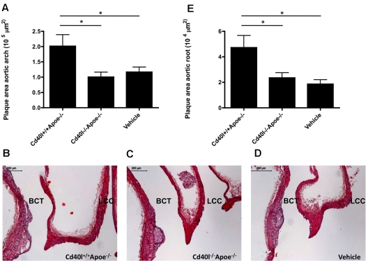
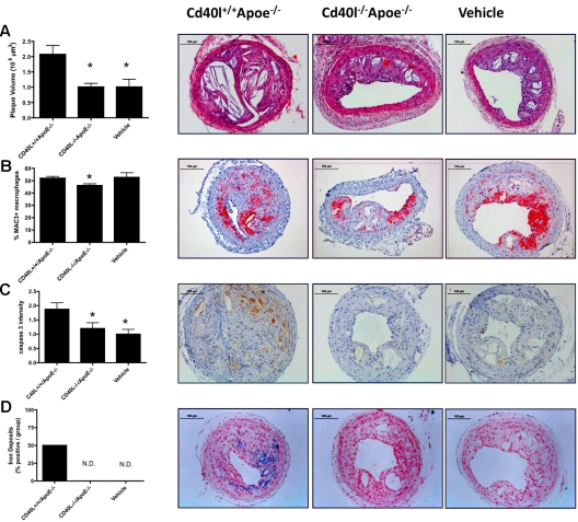
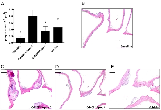
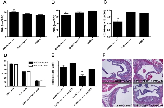
CD40 ligand (CD40L), identified as a costimulatory molecule expressed on T cells, is also expressed and functional on platelets. We investigated the thrombotic and inflammatory contributions of platelet CD40L in atherosclerosis. Although CD40L-deficient (Cd40l(-/-)) platelets exhibited impaired platelet aggregation and thrombus stability, the effects of platelet CD40L on inflammatory processes in atherosclerosis were more remarkable. Repeated injections of activated Cd40l(-/-) platelets into Apoe(-/-) mice strongly decreased both platelet and leukocyte adhesion to the endothelium and decreased plasma CCL2 levels compared with wild-type platelets. Moreover, Cd40l(-/-) platelets failed to form proinflammatory platelet-leukocyte aggregates. Expression of CD40L on platelets was required for platelet-induced atherosclerosis as injection of Cd40l(-/-) platelets in contrast to Cd40l(+/+) platelets did not promote lesion formation. Remarkably, injection of Cd40l(+/+), but not Cd40l(-/-), platelets transiently decreased the amount of regulatory T cells (Tregs) in blood and spleen. Depletion of Tregs in mice injected with activated Cd40l(-/-) platelets abrogated the athero-protective effect, indicating that CD40L on platelets mediates the reduction of Tregs leading to accelerated atherosclerosis. We conclude that platelet CD40L plays a pivotal role in atherosclerosis, not only by affecting platelet-platelet interactions but especially by activating leukocytes, thereby increasing platelet-leukocyte and leukocyte-endothelium interactions.


Figures

Figure 1
Figure 1
CD40L enhances collagen-dependent thrombus formation in Apoe-deficient mice under flow. (A) Heparin/D-phenylalanyl-L-prolyl-L-arginine chloromethylketone-anticoagulated blood from Cd40l+/+Apoe−/− or Cd40l−/−Apoe−/− mice was passed over VWF or collagen at shear rates of 1700 or 1000 seconds, respectively. (B) Representative phase-contrast images for platelets perfused over collagen are shown after 4 minutes of flow. Arrows indicate platelet aggregates (n = 4-6). *P < .05. (C) Size distribution of multiplatelet thrombi on collagen, established from morphometric image analysis reported as numbers of platelets per agglomerate. *P < .001. (D) Role of CD40L and CD40 in the formation of platelet leukocyte-aggregate mice. Numbers of leukocytes (cells/mm2) on immobilized Cd40l+/+Apoe−/− or Cd40l−/−Apoe−/− platelets (n = 6). *P < .05. (E) After in vitro coincubation, flow cytometric analysis of frequencies of CD41+CD11b+ cell aggregates, demonstrating the contribution of both platelet CD40L and leukocyte CD40 (n = 6). *P < .05 vs WT/WT.
Figure 2
Figure 2
Platelet CD40L promotes leukocyte adhesion to the atherosclerotic arterial wall. (A-D) Intravital microscopy of adhering, rhodamine-labeled leukocytes in carotid arteries of Apoe−/− mice treated with activated Cd40l+/+Apoe−/−, Cd40l−/−Apoe−/− platelets, or vehicle every 5 days for 10 weeks (n = 4-6). *P < .05. (E) Plasma CCL2 levels after repeated injection of Cd40l−/−Apoe−/− platelets into Apoe−/− mice compared with injection of Cd40l+/+Apoe−/− platelets and vehicle-treated mice (n = 9). *P < .05. (F) Double immunohistochemistry for FVIII (blue) and CCL2 (red) of a CD40l+/+Apoe−/− atherosclerotic lesion. (G) Double immunohistochemistry for Mac3 (blue) and CCL2 (red) of a CD40l+/+Apoe−/− atherosclerotic lesion. (H) Endothelial (luminal) deposition of CCL2, quantified by immunohistochemical staining, is decreased in the Cd40l−/−Apoe−/− treatment group compared with the Cd40l+/+Apoe−/− group (n = 6). *P < .05.
Figure 3
Figure 3
Adhesion of both platelets and leukocytes is impaired after injection of activated Cd40l−/−Apoe−/− platelets. Intravital microscopy of the endothelial wall of carotid arteries in 17-week-old Apoe−/− mice shows an impaired adhesion of intravenously administered calcein-labeled Cd40l−/−Apoe−/− platelets (green) (A). Subsequent rhodamine 6G injection shows a decreased adhesion of leukocytes (red) in Apoe−/− mice injected with Cd40l−/−Apoe−/− platelets compared with mice injected with Cd40l+/+Apoe−/− platelets (B) (n = 6). *P < .05. (C-D) Representative images of the external carotid artery. Scale bars represent 100 μm.
Figure 4
Figure 4
Platelet CD40L promotes atherosclerosis initiation. (A) Plaque area of the aortic arch, including the main branch points (brachiocephalic trunk [BCT], left common carotid artery [LCC], left subclavian artery) of Apoe−/− mice, injected every 5 days for 12 weeks with activated Cd40l+/+Apoe−/− or Cd40l−/−Apoe−/− platelets, or vehicle (n = 11/group). (B-D) Representative longitudinal sections (hematoxylin and eosin staining) of the aortic arch and main branch points. Note the early, foam cell-rich atherosclerotic lesions formed in the brachiocephalic trunk. Scale bar represents 200 μm. (E) Plaque area in the aortic root of the same mice (n = 9/group). *P < .05.
Figure 5
Figure 5
Platelet CD40L promotes advanced plaque development. Silastic collars were placed in 14-week-old Apoe−/− mice fed a 0.21% cholesterol diet. Activated Cd40l+/+Apoe−/− or Cd40l−/−Apoe−/− platelets, or vehicle were injected once every 5 days for 6 weeks. (A) Plaque volume was calculated from the plaque areas measured in 6 slices. (B) Macrophage infiltration: representative staining and percentage of Mac3+ cells of all plaque cells. (C) Cleaved caspase-3 was determined by immunohistochemistry and graded from 0 (not present) to 3 (highly present). (D) Iron deposits (percentage of mice containing lesions with iron depositions) were determined by Perl iron staining (n = 9/group); quantification (left) and representative figures (right) are shown. *P < .05 vs Cd40l+/+Apoe−/−. Scale bars represent 100 μm. N.D. indicates not detected.
Figure 6
Figure 6
Platelet CD40L contributes to the progression of established plaques. Seventeen-week-old Apoe−/− mice were injected with activated Cd40l+/+Apoe−/− or Cd40l−/−Apoe−/− platelets, or vehicle once every 5 days for 12 weeks. (A) Plaque area was quantified in the aortic arch and main branch points at 29 weeks and compared with the baseline lesion size of 17-week-old Apoe−/− mice. (B-E) Representative longitudinal sections (hematoxylin and eosin staining; n = 8/group). *P < .05 vs Cd40l+/+Apoe−/−. Scale bars represent 200 μm.
Figure 7
Figure 7
Platelet CD40L transiently disturbs T-cell homeostasis. Flow cytometric analysis of T-cell distribution in blood from adult Apoe−/− mice treated with activated Cd40l+/+Apoe−/− or Cd40l−/−Apoe−/− platelets, or vehicle 12 hours after injection. Percentage of CD4+ T cells (A), CD8+ T cells (B), and CD4+CD25+Foxp3+ regulatory T cells (C). (D) Forty-eight hours after injection of activated platelets, no differences in T-cell phenotype were observed. Anti-CD25 treatment increases atherosclerotic plaque area (E) on injection of Cd40l+/+Apoe−/− platelets and reverses the protective effect of platelet-CD40L deficiency on atherosclerosis. (E) Quantification and (F) representative figures of atherosclerotic plaques in the aortic root of Apoe−/− mice treated with activated Cd40l+/+Apoe−/− or Cd40l−/−Apoe−/− platelets in the absence or presence of an anti-CD25 antibody (n = 30). *P < .05 vs Cd40l+/+Apoe−/−.

Comment in

  • Platelets suppress Treg recruitment.
    Moura R, Tjwa M. Moura R, et al. Blood. 2010 Nov 18;116(20):4035-7. doi: 10.1182/blood-2010-09-303396. Blood. 2010. PMID: 21088138 No abstract available.

References

    1. Schonbeck U, Libby P. CD40 signaling and plaque instability. Circ Res. 2001;89(12):1092–1103. - PubMed
    1. Schonbeck U, Libby P. The CD40/CD154 receptor/ligand dyad. Cell Mol Life Sci. 2001;58(1):4–43. - PMC - PubMed
    1. Weber C, Zernecke A, Libby P. The multifaceted contributions of leukocyte subsets to atherosclerosis: lessons from mouse models. Nat Rev Immunol. 2008;8(10):802–815. - PubMed
    1. Galkina E, Ley K. Immune and inflammatory mechanisms of atherosclerosis. Annu Rev Immunol. 2009;27:165–197. - PMC - PubMed
    1. Mach F, Schonbeck U, Sukhova GK, Atkinson E, Libby P. Reduction of atherosclerosis in mice by inhibition of CD40 signalling. Nature. 1998;394(6689):200–203. - PubMed

Publication types

MeSH terms