. 2010 Oct 8;330(6001):231-5.
doi: 10.1126/science.1189435. Epub 2010 Aug 12.

Histone H3 Thr-3 phosphorylation by Haspin positions Aurora B at centromeres in mitosis

Affiliations

Histone H3 Thr-3 phosphorylation by Haspin positions Aurora B at centromeres in mitosis

Fangwei Wang et al. Science. .

Abstract

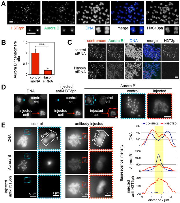
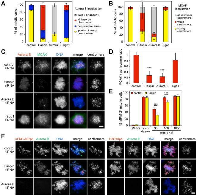
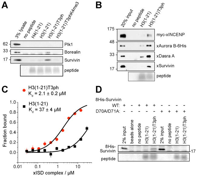
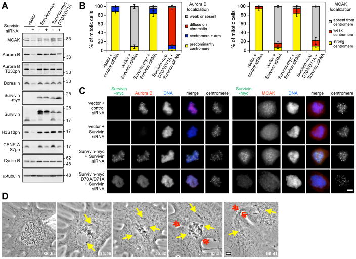
Aurora B is a component of the chromosomal passenger complex (CPC) required for correct spindle-kinetochore attachments during chromosome segregation and for cytokinesis. The chromatin factors that recruit the CPC to centromeres are unknown, however. Here we show that phosphorylation of histone H3 threonine 3 (H3T3ph) by Haspin is necessary for CPC accumulation at centromeres and that the CPC subunit Survivin binds directly to H3T3ph. A nonbinding Survivin-D70A/D71A mutant does not support centromeric CPC concentration, and both Haspin depletion and Survivin-D70A/D71A mutation diminish centromere localization of the kinesin MCAK and the mitotic checkpoint response to taxol. Survivin-D70A/D71A mutation and microinjection of H3T3ph-specific antibody both compromise centromeric Aurora B functions but do not prevent cytokinesis. Therefore, H3T3ph generated by Haspin positions the CPC at centromeres to regulate selected targets of Aurora B during mitosis.


Figures

Fig. 1
Fig. 1. Haspin RNAi or microinjection of anti-H3T3ph delocalizes Aurora-B from mitotic centromeres
(A) Immunofluorescence microscopy of nocodazole-arrested HeLa chromosome spreads reveals colocalization of H3T3ph and Aurora-B. (B, C) Cells were transfected with siRNA, and prepared as in (A). The Aurora-B/centromere autoantigen intensity ratio was determined by immunofluorescence at approximately 40 centromeres in 9 cells per condition (B). Means + SD are shown (N=3). *** p<0.001 by Student t test. Example images are shown in (C). (D, E) Nocodazole and MG132-treated LLC-PK cells were microinjected with anti-H3T3ph. After ~2 h, cells were subject to immunofluorescence staining for Aurora-B and with anti-rabbit antibodies to reveal microinjected antibody. Line scans of chromosomes are shown in (E). Yellow highlights the centromere. Scale bars = 5 μm unless noted.
Fig. 2
Fig. 2. Haspin RNAi delocalizes centromeric MCAK and compromises the spindle checkpoint, but has minor effects on Aurora-B activity toward CENP-AS7 and H3S10
(A, B) U2OS cells were transfected with siRNA, treated with nocodazole for 1–2 h, and subject to immunofluorescence staining. Approximately 100 mitotic cells in each condition were classified according to the localization of Aurora B (A) or MCAK (B). Means +/− SD are shown (N=3). (C) Example images of cells treated as above. (D) Following treatment as above, the MCAK/centromere autoantigen intensity ratio was determined at approximately 30 centromeres in 10–11 mitotic cells per condition. Means + SD are shown (N=3). (E) HeLa cells were transfected with siRNA, treated with thymidine for 24 h, and released into medium containing the compounds indicated. After 18 h, mitotic indices were determined by flow cytometry with MPM-2 antibody. Means + SD are shown (N=4). In (D and E), *** p<0.001 by Bonferroni’s multiple comparison test compared to corresponding control. (F) Immunofluorescence microscopy of CENP-AS7ph and H3S10ph in mitotic siRNA-transfected U2OS cells. Scale bars = 5 μm.
Fig. 3
Fig. 3. The CPC binds H3T3ph through Survivin
(A) Lysates of nocodazole-arrested HeLa cells were incubated with bead-immobilized histone peptides. Protein binding was visualized by immunoblotting, and peptide loading by Coomassie Blue staining. (B) Binding of recombinant Xenopus CPC to H3 peptides analyzed as above. (C) Binding of Xenopus ISD complex to H3 peptides measured by fluorescence anisotropy. Means +/− SD are shown (N=3). (D) Binding of recombinant human 8His-Survivin to H3 peptides visualized by Coomassie Blue staining.
Fig. 4
Fig. 4. CPC function at centromeres requires Survivin binding to H3T3ph
(A) Lysate immunoblotting of nocodazole-arrested HeLa cells stably expressing Survivin-myc or D70A/D71A mutant and transfected with siRNA as indicated. (B) The localization of Aurora-B and MCAK was classified by immunofluorescence microscopy in approximately 100 HeLa cells per condition. Means +/− SD are shown (N=3). (C) Example images of cells treated as in (B). (D) A Xenopus S3 cell microinjected with anti-H3T3ph exits mitosis in the presence of unaligned chromosomes (yellow arrows) with clear formation of a cleavage furrow (red arrows). Time is in min:s. Three of three injected S3 cells behaved similarly. See Movie S2. Scale bars = 5 μm.

Comment in

References

    1. Ruchaud S, Carmena M, Earnshaw WC. Nat Rev Mol Cell Biol. 2007;8:798. - PubMed
    1. Hsu JY, et al. Cell. 2000;102:279. - PubMed
    1. Andrews PD, et al. Dev Cell. 2004;6:253. - PubMed
    1. Lan W, et al. Curr Biol. 2004;14:273. - PubMed
    1. Zeitlin SG, Shelby RD, Sullivan KF. J Cell Biol. 2001;155:1147. - PMC - PubMed

Publication types

MeSH terms