. 2010 Sep 24;329(5999):1671-5.
doi: 10.1126/science.1189664. Epub 2010 Aug 12.

Dendritic discrimination of temporal input sequences in cortical neurons

Affiliations

Dendritic discrimination of temporal input sequences in cortical neurons

Tiago Branco et al. Science. .

Abstract

The detection and discrimination of temporal sequences is fundamental to brain function and underlies perception, cognition, and motor output. By applying patterned, two-photon glutamate uncaging, we found that single dendrites of cortical pyramidal neurons exhibit sensitivity to the sequence of synaptic activation. This sensitivity is encoded by both local dendritic calcium signals and somatic depolarization, leading to sequence-selective spike output. The mechanism involves dendritic impedance gradients and nonlinear synaptic N-methyl-D-aspartate receptor activation and is generalizable to dendrites in different neuronal types. This enables discrimination of patterns delivered to a single dendrite, as well as patterns distributed randomly across the dendritic tree. Pyramidal cell dendrites can thus act as processing compartments for the detection of synaptic sequences, thereby implementing a fundamental cortical computation.


Figures

Fig. 1
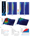
Fig. 1. Single dendrites are sensitive to the direction and velocity of synaptic input patterns.
(A) Layer 2/3 pyramidal cell filled with Alexa 594; yellow box indicates the selected dendrite. (B) Uncaging spots (yellow) along the selected dendrite. (C) Average individual uncaging responses at the soma. (D) Somatic responses to IN and OUT directions at 2.3 μm/ms (average in bold). (E) Comparing peak amplitudes for IN and OUT sequences at the optimal velocity for direction selectivity (green circle: example shown in D). (F) Direction-selective responses at different velocities. (G) Relationship between peak voltage and input velocity (values normalized to the maximum response in the IN direction for each cell, n = 15). (H) Relationship between direction selectivity and input velocity (n = 15). (I) Direction selectivity of spike probability; population data shown in J (P = 0.0013, n = 7). (K) Relationship between spike probability and velocity (n = 7, average of both directions).
Fig. 2
Fig. 2. Dendritic calcium influx is direction and velocity sensitive.
(A) Basal dendrite of a layer 2/3 pyramidal neuron; uncaging locations indicated in yellow, linescan profile used for Ca++ imaging in red. (B) Spatiotemporal profile of Ca++ signals triggered by IN and OUT input patterns at two different input velocities. (C) 3D plot of the data in B (2.3 μm/ms). (D) Relationship between Ca++ signals and input velocity (ΔF/F values normalized to the mean ΔF/F of all velocities in the IN direction of each cell). (E) Relationship between direction selectivity of Ca++ signals and input velocity. (F) Average spatial profile of the integrated Ca++ transient across the dendrite (n = 5 cells; lines indicate s.e.m.; bar indicates region of statistical significance).
Fig. 3
Fig. 3. NMDA receptor activation is required for robust velocity and direction coding.
(A) Somatic responses to IN and OUT sequences (coloured traces) and linear sum of the individual synaptic responses (gray traces). (B) Left, activating more synapses (1-7) produces EPSPs that are progressively larger than the linear sum of the individual responses. Right, summary plot of 8 cells. (C) As in A, but in the presence of AP5, which blocks the supralinearity. (D) Velocity and direction sensitivity is abolished in AP5. (E) Comparing peak amplitudes for IN and OUT sequences at the optimal velocity for direction selectivity (green circle: example shown in C; cf. Fig. 1E). (F) Summary plot of direction selectivity vs velocity (cf. Fig. 1G). (G) Reduced velocity sensitivity in AP5 (EPSPs normalized to the average IN maximum in control cells in the absence of APV; cf. Fig. 1H).
Fig. 4
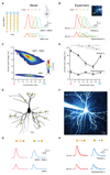
Fig. 4. Dendritic discrimination of complex input sequences.
(A) Random patterns of 9 inputs were generated and sorted by their directionality index (see Materials and Methods). Four sample patterns (numbers showing activation order) and corresponding directionality index are shown. Patterns were played into a single dendrite in a pyramidal cell model or B, experimentally in single pyramidal cell dendrites; peak response is different for different patterns, and responses become similar with hyperpolarization (or without NMDA receptors in model). (C) Pattern separability (measured by somatic peak EPSP distribution) was much greater with NMDA conductances present. (D) Peak EPSP voltage measured experimentally depends on input pattern similarly to the model (panel C); stars indicate significant differences between EPSP peaks for the example shown; hyperpolarization reduces the ability to discriminate different patterns. (E, F) Layer 2/3 pyramidal cell with uncaging spots (yellow) randomly distributed across 8 dendrites. Somatic responses to sequential uncaging in forward (red traces) or reverse sequence (blue traces; average in bold) are different at resting potential both experimentally (H) and in a compartmental model of the same neuron (G). Responses to the two sequences become similar upon hyperpolarization (H) or by removing synaptic NMDA receptors (G).

Comment in

Similar articles

Cited by

References

    1. Meister M, Lagnado L, Baylor DA. Science. 1995;270:1207. - PubMed
    1. deCharms RC, Merzenich MM. Nature. 1996;381:610. - PubMed
    1. Wehr M, Laurent G. Nature. 1996;384:162. - PubMed
    1. Johansson RS, Birznieks I. Nat Neurosci. 2004;7:170. - PubMed
    1. Thorpe S, Delorme A, Van Rullen R. Neural Netw. 2001;14:715. - PubMed

Publication types

MeSH terms