. 2010 Nov;299(5):G1118-27.
doi: 10.1152/ajpgi.00131.2010. Epub 2010 Aug 12.

Bifidobacterium bifidum reduces apoptosis in the intestinal epithelium in necrotizing enterocolitis

Affiliations

Bifidobacterium bifidum reduces apoptosis in the intestinal epithelium in necrotizing enterocolitis

Ludmila Khailova et al. Am J Physiol Gastrointest Liver Physiol. 2010 Nov.

Abstract

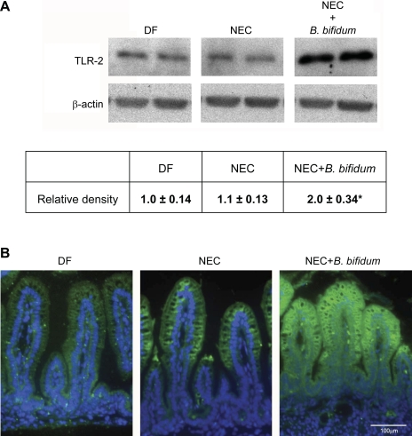
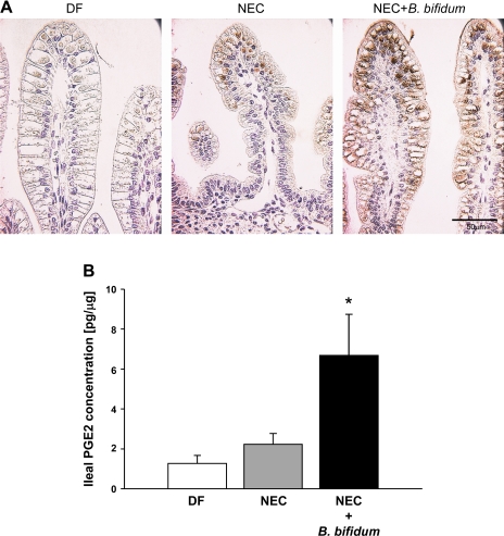
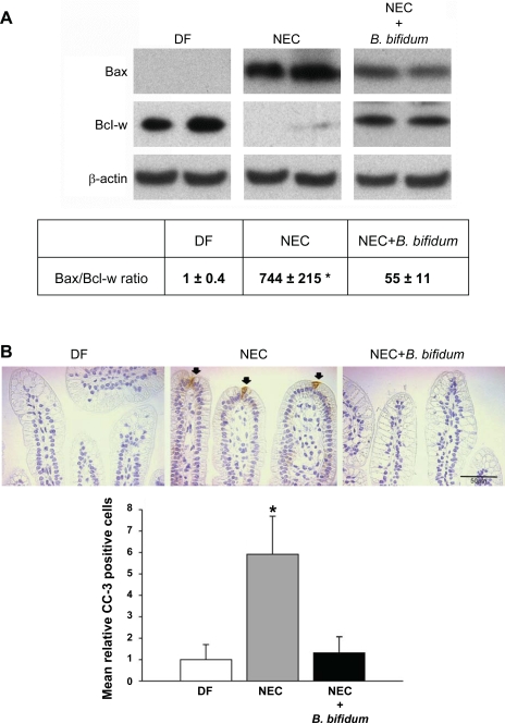
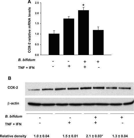
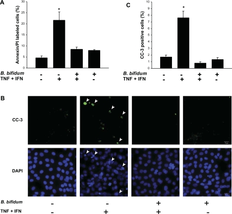
Necrotizing enterocolitis (NEC) is a devastating intestinal disease of neonates, and clinical studies suggest the beneficial effect of probiotics in NEC prevention. Recently, we have shown that administration of Bifidobacterium bifidum protects against NEC in a rat model. Intestinal apoptosis can be suppressed by activation of cyclooxygenase-2 (COX-2) and increased production of prostaglandin E(2) (PGE(2)). The present study investigates the effect of B. bifidum on intestinal apoptosis in the rat NEC model and in an intestinal epithelial cell line (IEC-6), as a mechanism of protection against mucosal injury. Premature rats were divided into the following three groups: dam fed, hand fed with formula (NEC), or hand fed with formula supplemented with B. bifidum (NEC + B. bifidum). Intestinal Toll-like receptor-2 (TLR-2), COX-2, PGE(2), and apoptotic regulators were measured. The effect of B. bifidum was verified in IEC-6 cells using a model of cytokine-induced apoptosis. Administration of B. bifidum increased expression of TLR-2, COX-2, and PGE(2) and significantly reduced apoptosis in the intestinal epithelium of both in vivo and in vitro models. The Bax-to-Bcl-w ratio was shifted toward cell survival, and the number of cleaved caspase-3 positive cells was markedly decreased in B. bifidum-treated rats. Experiments in IEC-6 cells showed anti-apoptotic effect of B. bifidum. Inhibition of COX-2 signaling blocked the protective effect of B. bifidum treatment in both in vivo and in vitro models. In conclusion, oral administration of B. bifidum activates TLR-2 in the intestinal epithelium. B. bifidum increases expression of COX-2, which leads to higher production of PGE(2) in the ileum and protects against intestinal apoptosis associated with NEC. This study indicates the ability of B. bifidum to downregulate apoptosis in the rat NEC model and in IEC-6 cells by a COX-2-dependent matter and suggests a molecular mechanism by which this probiotic reduces mucosal injury and preserves intestinal integrity.


Figures

Fig. 1.
Fig. 1.
Effect of oral administration of Bifidobacterium bifidum on expression and localization of Toll-like receptor-2 (TLR-2) in the ileum. A: representative TLR-2 (90-kDa) bands from Western blot analyses are shown for dam-fed littermates fed by surrogate mothers as a baseline control (DF, n = 5), littermates hand fed with formula (NEC, n = 5), and littermates hand fed with formula containing 5 × 10⁶ colony-forming units (CFU) of B. bifidum OLB6378 in two feedings per day (NEC + B. bifidum, n = 5). All samples were analyzed on the same gel. Mean relative density to β-actin was calculated for all groups. *P ≤ 0.05 vs. NEC and DF. B: representative slides from DF, NEC, and NEC + B. bifidum groups (n = 6 animals/experimental group). Magnification: ×400.
Fig. 2.
Fig. 2.
Effect of B. bifidum treatment on cyclooxygenase-2 (COX-2) expression and prostaglandin E2 (PGE2) production in the ileal tissue. A: ileal tissue stained for COX-2; representative slides from DF, NEC, and NEC + B. bifidum groups are shown (n = 6 animals/experimental group). Magnification: ×400. B: PGE2 concentration (pg/ml) in ileal homogenates of DF (n = 7), NEC (n = 7), and B. bifidum-treated animals (n = 7) calculated per 1 μg of protein. *P ≤ 0.05 vs. NEC and DF.
Fig. 3.
Fig. 3.
Evaluation of apoptotic markers in the ileum after B. bifidum treatment of NEC. A: representative Bax (26 kDa) and Bcl-w (21 kDa) bands from Western blot analyses of ileal tissue from DF (n = 5), NEC (n = 5), and NEC + B. bifidum (n = 5) groups and the ratio between pro-apoptotic Bax and anti-apoptotic Bcl-w. All samples were analyzed on the same gel. *P ≤ 0.05 vs. NEC + B. bifidum and DF. B: representative slides from DF, NEC, and NEC + B. bifidum groups stained for cleaved caspase-3 (CC-3). Arrows indicate CC-3 positively stained cells. Magnification: ×400. Mean relative number of CC-3 positive cells in the site of NEC injury (n = 5 animals/experimental group). Data are expressed as mean CC-3 positive cells/100 epithelial cells. *P ≤ 0.05 vs. NEC + B. bifidum and DF.
Fig. 4.
Fig. 4.
TLR-2 expression in IEC-6 cells evaluated by fluorescent microscopy. Cells were treated with or without tumor necrosis factor (TNF)-α and interferon (IFN)-γ (400 ng/ml each) for 4 h and with or without a 15-min pretreatment with B. bifidum. Arrowheads indicate representative TLR-2-expressing cells and their nuclei [stained with 4′,6-diamidino-2-phenylindole (DAPI)].
Fig. 5.
Fig. 5.
Evaluation of COX-2 in IEC-6 cells. Cells were treated with or without TNF-α and IFN-γ (400 ng/ml each) for 4 h with or without pretreatment for 15 min with B. bifidum. A: COX-2 mRNA levels evaluated using real-time PCR. The mean steady-state mRNA level for the control group (no treatment) was assigned a value of 1.0, and mean mRNA levels for all other groups were determined relative to this number. *P ≤ 0.05 vs. control (no treatment) and B. bifidum (+) TNF + INF (−). B: representative COX-2 (70-kDa) bands from Western blot analyses of IEC-6 cell lysates. Mean relative density to β-actin is shown for all groups. *P ≤ 0.05 vs. control (no treatment); B. bifidum (−) TNF + INF (+); and B. bifidum (+) TNF + INF (−).
Fig. 6.
Fig. 6.
Apoptosis in IEC-6 cells and the effect of B. bifidum pretreatment. Cells were treated with or without TNF-α and IFN-γ (400 ng/ml each) for 4 h and with or without a 15-min pretreatment with B. bifidum. A: percentage of annexin/propidium iodide (PI)-labeled cells analyzed by flow cytometry. *P ≤ 0.05 vs. control (no treatment); B. bifidum (+) TNF + INF (+); and B. bifidum (+) TNF + INF (−). B: CC-3-stained cells evaluated by fluorescent microscopy. Arrowheads indicate representative apoptotic cells/nuclei. Magnification: ×400. C: the percentage of CC-3-positive cells. *P ≤ 0.05 vs. control (no treatment); B. bifidum (+) TNF + INF (+); and B. bifidum (+) TNF + INF (−).

References

    1. Bin-Nun A, Bromiker R, Wilschanski M, Kaplan M, Rudensky B, Caplan M, Hammerman C. Oral probiotics prevent necrotizing enterocolitis in very low birth weight neonates. J Pediatr 147: 192–196, 2005 - PubMed
    1. Bos J, Smithee L, McClane B, Distefano RF, Uzal F, Songer JG, Mallonee S, Crutcher JM. Fatal necrotizing colitis following a foodborne outbreak of enterotoxigenic Clostridium perfringens type A infection. Clin Infect Dis 40: e78–e83, 2005 - PubMed
    1. Cario E, Gerken G, Podolsky DK. Toll-like receptor 2 controls mucosal inflammation by regulating epithelial barrier function. Gastroenterology 132: 1359–1374, 2007 - PubMed
    1. Cario E, Gerken G, Podolsky DK. Toll-like receptor 2 enhances ZO-1-associated intestinal epithelial barrier integrity via protein kinase C. Gastroenterology 127: 224–238, 2004 - PubMed
    1. Chang YJ, Wu MS, Lin JT, Sheu BS, Muta T, Inoue H, Chen CC. Induction of cyclooxygenase-2 overexpression in human gastric epithelial cells by Helicobacter pylori involves TLR2/TLR9 and c-Src-dependent nuclear factor-kappaB activation. Mol Pharmacol 66: 1465–1477, 2004 - PubMed

Publication types

MeSH terms