. 2010 Dec;299(6):G1308-18.
doi: 10.1152/ajpgi.00156.2010. Epub 2010 Aug 12.

Vagal nerve stimulation protects against burn-induced intestinal injury through activation of enteric glia cells

Affiliations

Vagal nerve stimulation protects against burn-induced intestinal injury through activation of enteric glia cells

Todd W Costantini et al. Am J Physiol Gastrointest Liver Physiol. 2010 Dec.

Abstract

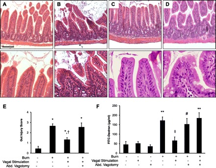
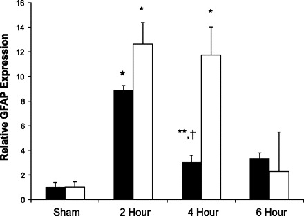
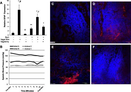
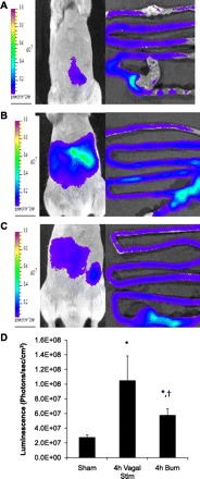
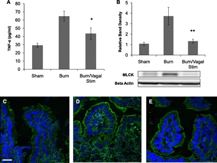
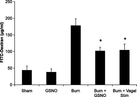
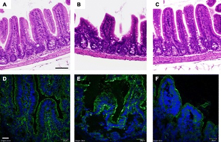
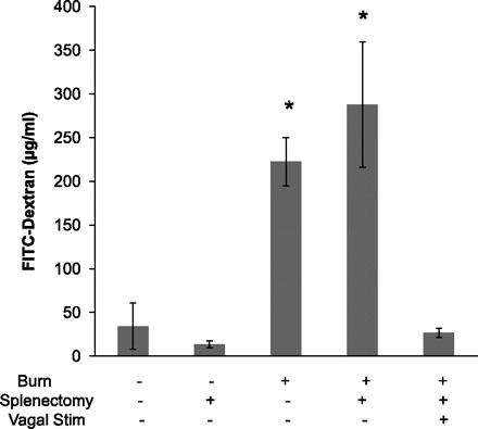
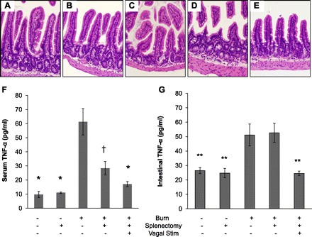
The enteric nervous system may have an important role in modulating gastrointestinal barrier response to disease through activation of enteric glia cells. In vitro studies have shown that enteric glia activation improves intestinal epithelial barrier function by altering the expression of tight junction proteins. We hypothesized that severe injury would increase expression of glial fibrillary acidic protein (GFAP), a marker of enteric glial activation. We also sought to define the effects of vagal nerve stimulation on enteric glia activation and intestinal barrier function using a model of systemic injury and local gut mucosal involvement. Mice with 30% total body surface area steam burn were used as model of severe injury. Vagal nerve stimulation was performed to assess the role of parasympathetic signaling on enteric glia activation. In vivo intestinal permeability was measured to assess barrier function. Intestine was collected to investigate changes in histology; GFAP expression was assessed by quantitative PCR, by confocal microscopy, and in GFAP-luciferase transgenic mice. Stimulation of the vagus nerve prevented injury-induced intestinal barrier injury. Intestinal GFAP expression increased at early time points following burn and returned to baseline by 24 h after injury. Vagal nerve stimulation prior to injury increased GFAP expression to a greater degree than burn alone. Gastrointestinal bioluminescence was imaged in GFAP-luciferase transgenic animals following either severe burn or vagal stimulation and confirmed the increased expression of intestinal GFAP. Injection of S-nitrosoglutathione, a signaling molecule released by activated enteric glia cells, following burn exerts protective effects similar to vagal nerve stimulation. Intestinal expression of GFAP increases following severe burn injury. Stimulation of the vagus nerve increases enteric glia activation, which is associated with improved intestinal barrier function. The vagus nerve may mediate the signaling that occurs from the central nervous system to the enteric nervous system following gastrointestinal injury.


Figures

Fig. 1.
Fig. 1.
Vagal nerve stimulation attenuates burn-induced intestinal injury. Distal small intestine harvested from animals 4 h following 30% total body surface area (TBSA) burn (n = 3 animals per group). Sections of small intestine were stained with hematoxylin and eosin. High-power images of the villous tips from each section are shown below each image. A: section of normal small intestine from sham animal. B: intestine of burned animals displays evidence of histological pathology characterized by blunting of villi. C: gut sections harvested from animals that underwent cervical vagal nerve stimulation prior to severe burn. D: histological gut injury noted in animals that underwent abdominal vagotomy prior to stimulation of the vagus nerve. Size bar = 100 μm. E: intestinal injury was scored on a scale of 0 (normal) to 4 (severe) by a pathologist blinded to the experimental conditions. *P < 0.05 vs. Sham, †P = 0.05 vs. Burn and Burn/Vagotomy/Vagal Stimulation using the Kruskal-Wallis test followed by pairwise Mann-Whitney test. F: vagal nerve stimulation limits intestinal barrier dysfunction following injury (n = 4 animals per group). Vagal nerve stimulation prevents intestinal barrier dysfunction 4 h following 30% TBSA burn. Dividing the abdominal (Abd.) vagus nerve abrogates the protective effects of vagal nerve stimulation. Data are expressed as mean systemic FITC-dextran concentration ± SE. **P < 0.01 vs. Sham, #P < 0.05 vs. Sham, ‡P < 0.05 vs. Burn and Burn/Vagotomy/Vagal Stimulation by analysis of variance.
Fig. 2.
Fig. 2.
Severe burn increases expression of intestinal glial fibrillary acidic protein (GFAP). Quantitative PCR (n ≥ 3 animals per group) was performed on intestinal extracts obtained at several time points following burn (open bars) or cervical vagal nerve stimulation (solid bars). Activation of enteric glia cells occurs at early time points following burn, with an 8-fold increase in gut GFAP mRNA expression at 2 h following injury. Vagal nerve stimulation also causes activation of enteric glia cells. Intestinal GFAP mRNA expression increased by nearly 12-fold at 2 and 4 h following vagal nerve stimulation. Data are expressed as GFAP mRNA expression relative to sham ± SE. *P < 0.01 vs. Sham, **P < 0.05 vs. Sham, †P < 0.05 vs. 2-h burn using analysis of variance.
Fig. 3.
Fig. 3.
Vagal nerve stimulation alters intestinal GFAP expression. A: quantitative PCR was performed on intestinal extracts obtained 4 h following injury. Results are expressed as intestinal GFAP mRNA expression relative to sham ± SE. Vagal nerve stimulation (Stim) prior to severe burn results in augmented intestinal GFAP mRNA expression over animals subjected to burn alone. Performing a surgical abdominal vagotomy prior to cervical vagal nerve stimulation and subsequent severe burn abrogates the ability of vagal nerve stimulation to increase intestinal GFAP mRNA expression. B: arterial blood pressure was measured during vagal nerve stimulation to confirm that cervical vagal nerve stimulation did not cause hemodynamically significant changes in blood pressure. There was no significant change in arterial blood pressure during or immediately following vagal nerve stimulation. Intestinal GFAP+ enteric glia localization was assessed by confocal microscopy of intestinal segments stained for GFAP (red) and 4,6-diamidino-2-phenylindole (DAPI; blue). C: there is some background GFAP expression in sham animals. D: GFAP staining is seen extending toward the intestinal villi in sections from animals 4 h following burn. E: vagal nerve stimulation prior to burn also results in increased staining for GFAP compared with sham. F: abdominal vagotomy prior to vagal nerve stimulation and burn results in a pattern of staining similar to sham, suggesting the importance of vagal nerve signaling in enteric glia activation. *P < 0.01 vs. Sham, **P < 0.05 vs. Sham, †P < 0.02 vs. Burn, #P < 0.02 vs. Burn and Burn/Vagotomy/Vagal Stimulation, ‡P < 0.001 vs. all groups except Sham by analysis of variance. Size bar = 20 μm.
Fig. 4.
Fig. 4.
In vivo increases in abdominal luminescence in GFAP-luc mice. Transgenic mice expressing luciferase under control of the GFAP promoter were imaged in a Xenogen IVIS Lumina 2 h following burn (n > 3 animals per group). A: representative sham GFAP-luc mouse demonstrating background GFAP expression with resected intestine from confirming luminescence from the intestine. B: in vivo luminescence from the gut of GFAP-luc mice is elevated 4 h following stimulation of the cervical vagus nerve. C: increased luminescence is also seen from the abdomen of GFAP-luc mice 4 h following burn. D: quantification of luminescence measured from the abdomen of all GFAP-luc animals imaged, *P < 0.05 vs. Sham, †P < 0.05 vs. Vagal Nerve Stimulation.
Fig. 5.
Fig. 5.
Vagal nerve stimulation prevents burn-induced intestinal barrier injury. A: intestinal TNF-α levels measured from distal ileum harvested 4 h following injury using ELISA. Vagal nerve stimulation prevents the increase in TNF-α seen following burn injury, *P < 0.05 vs. Burn. B: changes in intestinal myosin light chain kinase (MLCK) expression 4 h following burn by Western blot. There is nearly a 4-fold increase in intestinal MLCK expression following burn. Animals undergoing vagal nerve stimulation exhibited intestinal MLCK expression similar to sham, **P < 0.05 vs. Burn by analysis of variance. Intestinal segments were harvested 4 h following burn and stained for phosphorylated myosin light chain (MLC, green) and DAPI (blue) by confocal microscopy. C: minimal staining seen in sham animals. D: staining for phosphorylated MLC is increased following burn. E: vagal nerve stimulation prior to burn decreases staining for phosphorylated MLC. Bar = 20 μm.
Fig. 6.
Fig. 6.
Postinjury vagal nerve stimulation protects against gut barrier injury. In vivo intestinal permeability to 4-kDa FITC-dextran was measured 4 h following in burn injury. Animals underwent either postinjury vagal nerve stimulation or S-nitrosoglutathione (GSNO) injection. Stimulation of the vagus nerve 4 h following burn injury decreases intestinal permeability compared with sham. GSNO injection also modulates gut barrier function following severe burn, attenuating burn-induced intestinal permeability. *P < 0.03 vs. Burn.
Fig. 7.
Fig. 7.
Injection of GSNO decreases intestinal injury following severe burn. Intestinal injury was assessed by histology. B: sham. B: there is clear evidence of gut injury in sections obtained from animals subjected to severe burn. C: sections of intestine from burned animals injected with GSNO show minimal evidence of injury, similar to sham. Black size bar = 100 μm. Changes in phosphorylated MLC (green) were assessed by confocal microscopy. Cell nuclei were stained with DAPI (blue). D: there is minimal staining for phosphorylated MLC seen in sham animals. E: staining for phosphorylated MLC is increased following burn. F: injection of GSNO prior to burn decreases staining for phosphorylated MLC. White size bar = 30 μm.
Fig. 8.
Fig. 8.
Vagal nerve stimulation prevents burn-induced intestinal permeability in splenectomized animal. In vivo intestinal permeability to 4-kDa FITC-dextran was measured 4 h following injury. Increased intestinal permeability was seen in animals that underwent splenectomy immediately prior to burn injury. The protective effects of vagal nerve stimulation are seen in burned animals that underwent splenectomy. Taken together, this suggests the gut-protective effects of vagal nerve stimulation are not due to modulation of splenic cytokine production. *P < 0.02 vs. Sham, Splenectomy, and Burn/Splenectomy/Vagal Stim groups.
Fig. 9.
Fig. 9.
Gut-protective effects of vagal nerve stimulation are independent of changes in circulating TNF-α. Distal small intestine harvested from animals 4 h following injury (n = 3 animals per group). Sections of small intestine stained with hematoxylin and eosin. Representative sections are shown of normal small intestine from sham (A) and following splenectomy (B). Intestines of a representative animal following burn (C) and an animal that underwent splenectomy prior to burn (D) display evidence of histological gut injury. E: a normal gut section harvested from a representative animal that underwent splenectomy prior to vagal nerve stimulation and severe burn. F: serum TNF-α was measured 4 h following injury using ELISA. There is a decrease in circulating TNF-α in animals that underwent splenectomy. *P < 0.001 vs. Burn, †P < 0.01 vs. Burn. G: TNF-α measured from intestinal extracts obtained 4 h following burn injury. There is evidence of local intestinal TNF-α production that correlates with the intestinal injury seen by intestinal permeability and histology. **P < 0.05 vs. Burn and Burn/Splenectomy.

Similar articles

Cited by

References

    1. Al-Sadi R, Boivin M, Ma T. Mechanism of cytokine modulation of epithelial tight junction barrier. Front Biosci 14: 2765–2778, 2009 - PMC - PubMed
    1. Bansal V, Costantini T, Kroll L, Peterson C, Loomis W, Eliceiri B, Baird A, Wolf P, Coimbra R. Traumatic brain injury and intestinal dysfunction: uncovering the neuro-enteric axis. J Neurotrauma 26: 1353–1359, 2009 - PMC - PubMed
    1. Blikslager AT, Moeser AJ, Gookin JL, Jones SL, Odle J. Restoration of barrier function in injured intestinal mucosa. Physiol Rev 87: 545–564, 2007 - PubMed
    1. Bradley JS, Jr, Parr EJ, Sharkey KA. Effects of inflammation on cell proliferation in the myenteric plexus of the guinea-pig ileum. Cell Tissue Res 289: 455–461, 1997 - PubMed
    1. Bush TG, Savidge TC, Freeman TC, Cox HJ, Campbell EA, Mucke L, Johnson MH, Sofroniew MV. Fulminant jejuno-ileitis following ablation of enteric glia in adult transgenic mice. Cell 93: 189–201, 1998 - PubMed

Substances