

Different oxidative stress response in keratinocytes and fibroblasts of reconstructed skin exposed to non extreme daily-ultraviolet radiation
- PMID: 20706594
- PMCID: PMC2919404
- DOI: 10.1371/journal.pone.0012059
Different oxidative stress response in keratinocytes and fibroblasts of reconstructed skin exposed to non extreme daily-ultraviolet radiation
Abstract
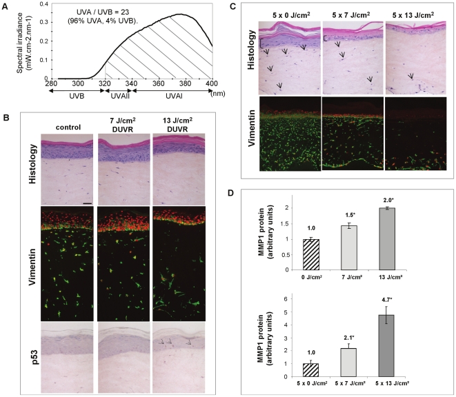
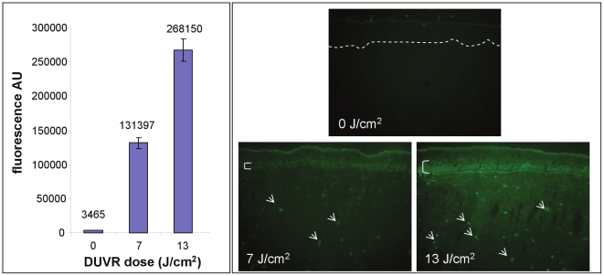
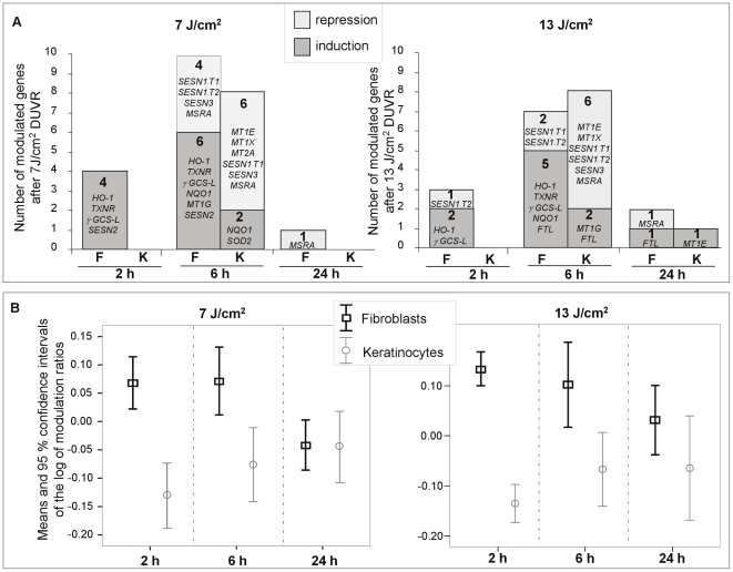
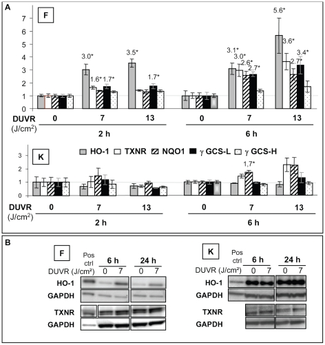
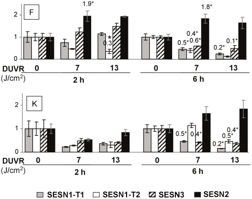
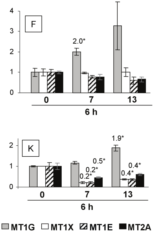
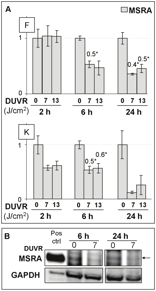
Experiments characterizing the biological effects of sun exposure have usually involved solar simulators. However, they addressed the worst case scenario i.e. zenithal sun, rarely found in common outdoor activities. A non-extreme ultraviolet radiation (UV) spectrum referred as "daily UV radiation" (DUVR) with a higher UVA (320-400 nm) to UVB (280-320 nm) irradiance ratio has therefore been defined. In this study, the biological impact of an acute exposure to low physiological doses of DUVR (corresponding to 10 and 20% of the dose received per day in Paris mid-April) on a 3 dimensional reconstructed skin model, was analysed. In such conditions, epidermal and dermal morphological alterations could only be detected after the highest dose of DUVR. We then focused on oxidative stress response induced by DUVR, by analyzing the modulation of mRNA level of 24 markers in parallel in fibroblasts and keratinocytes. DUVR significantly modulated mRNA levels of these markers in both cell types. A cell type differential response was noticed: it was faster in fibroblasts, with a majority of inductions and high levels of modulation in contrast to keratinocyte response. Our results thus revealed a higher sensitivity in response to oxidative stress of dermal fibroblasts although located deeper in the skin, giving new insights into the skin biological events occurring in everyday UV exposure.
Conflict of interest statement
Figures
Similar articles
-
[Human skin reconstructed in vitro as a model to study the keratinocyte, the fibroblast and their interactions: photodamage and repair processes].J Soc Biol. 2005;199(4):313-20. doi: 10.1051/jbio:2005032. J Soc Biol. 2005. PMID: 16738525 Review. French.
-
Modulations of gene expression induced by daily ultraviolet light can be prevented by a broad spectrum sunscreen.J Photochem Photobiol B. 2012 Nov 5;116:37-47. doi: 10.1016/j.jphotobiol.2012.08.001. Epub 2012 Aug 13. J Photochem Photobiol B. 2012. PMID: 22960577
-
IL-1 receptor antagonist attenuates MAP kinase/AP-1 activation and MMP1 expression in UVA-irradiated human fibroblasts induced by culture medium from UVB-irradiated human skin keratinocytes.Int J Mol Med. 2005 Dec;16(6):1117-24. Int J Mol Med. 2005. PMID: 16273295
-
In vitro tools for photobiological testing: molecular responses to simulated solar UV of keratinocytes growing as monolayers or as part of reconstructed skin.Photochem Photobiol Sci. 2010 Apr;9(4):448-58. doi: 10.1039/b9pp00145j. Photochem Photobiol Sci. 2010. PMID: 20354637
-
Effects and interactions of increased environmental temperature and UV radiation on photoageing and photocarcinogenesis of the skin.Exp Dermatol. 2019 Feb;28 Suppl 1:23-27. doi: 10.1111/exd.13818. Exp Dermatol. 2019. PMID: 30698877 Review.
Cited by
-
Oxidative Stress as an Important Contributor to the Pathogenesis of Psoriasis.Int J Mol Sci. 2020 Aug 27;21(17):6206. doi: 10.3390/ijms21176206. Int J Mol Sci. 2020. PMID: 32867343 Free PMC article. Review.
-
Integrated transcriptome study of the tumor microenvironment for treatment response prediction in male predominant hypopharyngeal carcinoma.Nat Commun. 2023 Mar 16;14(1):1466. doi: 10.1038/s41467-023-37159-8. Nat Commun. 2023. PMID: 36928331 Free PMC article.
-
Exposure to non-extreme solar UV daylight: spectral characterization, effects on skin and photoprotection.Int J Mol Sci. 2014 Dec 23;16(1):68-90. doi: 10.3390/ijms16010068. Int J Mol Sci. 2014. PMID: 25546388 Free PMC article. Review.
-
Effects of Natural Antioxidants on Phospholipid and Ceramide Profiles of 3D-Cultured Skin Fibroblasts Exposed to UVA or UVB Radiation.Antioxidants (Basel). 2021 Apr 8;10(4):578. doi: 10.3390/antiox10040578. Antioxidants (Basel). 2021. PMID: 33918064 Free PMC article.
-
Diversity of biological effects induced by longwave UVA rays (UVA1) in reconstructed skin.PLoS One. 2014 Aug 20;9(8):e105263. doi: 10.1371/journal.pone.0105263. eCollection 2014. PLoS One. 2014. PMID: 25140898 Free PMC article.
References
-
- Gilchrest BA. Dermatoheliosis (Sun-induced aging) In: Gilchrest BA, editor. Photodamage. Cambridge USA: Blackwell Science Inc; 1995. pp. 97–116.
-
- Colipa . Brussels, Belgium: The European Cosmetic, Toiletry and Perfumary Association; 1994. Colipa sun protection factor test method.
-
- Sheehan JM, Young AR. The sunburn cell revisited: an update on mechanistic aspects. Photochem Photobiol Sci. 2002;1:365–377. - PubMed
-
- Young AR, Orchard GE, Harrison GI, Klock JL. The detrimental effects of daily sub-erythemal exposure on human skin in vivo can be prevented by a daily-care broad-spectrum sunscreen. J Invest Dermatol. 2007;127:975–978. - PubMed
Publication types
MeSH terms
Substances
LinkOut - more resources
Full Text Sources
Other Literature Sources
Medical
Molecular Biology Databases
Miscellaneous
