. 2010:2010:753512.
doi: 10.1155/2010/753512. Epub 2010 Jul 14.

Global distribution, public health and clinical impact of the protozoan pathogen cryptosporidium

Affiliations

Global distribution, public health and clinical impact of the protozoan pathogen cryptosporidium

Lorenza Putignani et al. Interdiscip Perspect Infect Dis. 2010.

Abstract

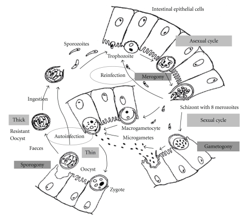
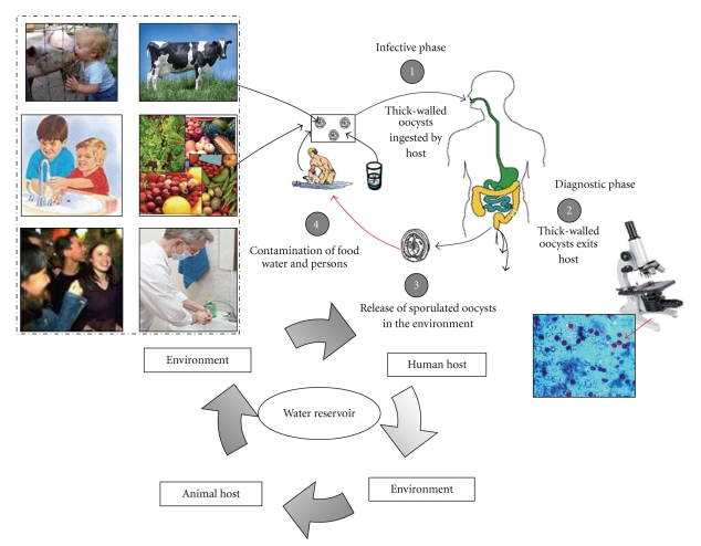
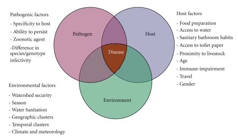
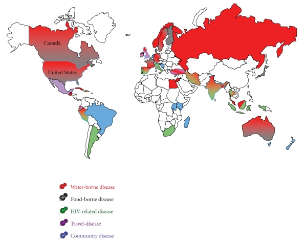
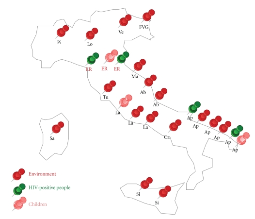
Cryptosporidium spp. are coccidians, oocysts-forming apicomplexan protozoa, which complete their life cycle both in humans and animals, through zoonotic and anthroponotic transmission, causing cryptosporidiosis. The global burden of this disease is still underascertained, due to a conundrum transmission modality, only partially unveiled, and on a plethora of detection systems still inadequate or only partially applied for worldwide surveillance. In children, cryptosporidiosis encumber is even less recorded and often misidentified due to physiological reasons such as early-age unpaired immunological response. Furthermore, malnutrition in underdeveloped countries or clinical underestimation of protozoan etiology in developed countries contribute to the underestimation of the worldwide burden. Principal key indicators of the parasite distribution were associated to environmental (e.g., geographic and temporal clusters, etc.) and host determinants of the infection (e.g., age, immunological status, travels, community behaviours). The distribution was geographically mapped to provide an updated picture of the global parasite ecosystems. The present paper aims to provide, by a critical analysis of existing literature, a link between observational epidemiological records and new insights on public health, and diagnostic and clinical impact of cryptosporidiosis.


Figures

Figure 1
Figure 1
Life cycle of Cryptosporidium in the enterocyte. Following oocyst ingestion by a host, and excystation, the sporozoites are released and parasitize epithelial cells of the gastrointestinal tract. In these cells, the parasites undergo asexual multiplication (schizogony or merogony) and then sexual multiplication (gametogony), producing microgamonts and macrogamonts. Upon fertilization of the macrogamonts by the microgametes, oocysts develop and sporulate in the infected host. Two different types of oocysts are produced: the thick-walled, which is commonly excreted by the host, after sporogony, and the thin-walled oocyst, which is primarily involved in autoinfection. Putignani and Menchella, 2010.
Figure 2
Figure 2
Description of transmission modes of Cryptosporidium. Following ingestion (and possibly inhalation) by a suitable host (e.g., human host), excystation occurs (infective stage, (1)). The released sporozoites invade epithelial cells of the gastrointestinal tract or other tissues, complete their cycle producing oocysts which exit host (diagnostic stage, (2)) and are released in the environment (3). Transmission of Cryptosporidium mainly occurs by ingestion of contaminated water (e.g., surface, drinking or recreational water), food sources (e.g., chicken salad, fruits, vegetables) or by person-to-person contact (community and hospital infections) (4). Zoonotic transmission of C. parvum occurs through exposure to infected animals (person-to-animal contact) or exposure to water (reservoir) contaminated by feces of infected animals (4). Putignani and Menchella, 2010.
Figure 3
Figure 3
Venn Diagram of factors leading to Cryptosporidium infection. Parasite, host and environmental indexes acting as key factors for the global burden of cryptosporidiosis. For details see Table 1. Putignani and Menchella, 2010.
Figure 4
Figure 4
Geography of worldwide occurrence of human cryptosporidiosis outbreaks and sporadic cases. A color-coded distribution of the main cases of cryptosporidosis reported in the literature during the last decade (1998–2008) for the entire population (adults and children) is here represented. Waterborne and foodborne diseases are represented by red and grey color, respectively. Spreading of the infection due to HIV immunological impairment is represented by green and travel-related disease by pink color. When not applicable the definition of waterborne and foodborne disease, the term community disease has been applied to person-to-person contacts and represented by a pale blue color. For countries characterised by two or three coexisting transmission modes, a double color-filling effect plus thick border lines have been used, consistently with the above reported code. Putignani and Menchella, 2010.
Figure 5
Figure 5
Geographical distribution of Italian studies on Cryptosporidium surveillance. A map of the principal surveillance studies performed on environmental and human samples is here reported by using the following color codes: red for environmental samples (water, animal); green for human samples associated to HIV in adults; pink for children samples. Symbols refer to different Italian regions: Ab, Abruzzo; Ap, Apulia; Ca, Campania; ER, Emilia Romagna; FVG, Friuli Venezia Giulia; La, Latium; Lo, Lombardia; Ma, Marche; Pi, Piedmont; Sa, Sardinia; Si, Sicily; Tu, Tuscany; Ve, Veneto. Putignani and Menchella, 2010.
Figure 6
Figure 6
Schematic life cycle of the most recurrent Cryptosporidium coinfective and coemerging parasitic agents. Panel (a). Giardia intestinalis (also called Giardia lamblia or Giardia duodenalis) is a flagellate parasite (Diplomonadida). Both cysts and trophozoites can be found in nondiarrheal feces (diagnostic and infective stages, (1)). Cysts are resistant forms and are responsible for transmission of giardiasis (infective stage, (2)). Infection occurs by the ingestion of water or food contaminated by cysts, or by the fecal-oral route (hands or fomites). In the small intestine, excystation releases trophozoites which multiply by longitudinal binary fission, remaining in the lumen of the proximal small bowel where they can be free or attached to the mucosa. Encystation occurs as the parasites transit toward the colon. Panel (b). Cyclospora cayetanensis is a coccidian parasite (Apicomplexa). When passed in stools, the oocyst is not infective (on the contrary of Cryptosporidium, thus direct fecal-oral transmission cannot occur). In the environment, sporulation occurs after days or weeks, resulting in division of the sporont into two sporocysts, each containing two sporozoites (diagnostic and infective stages, (1)). Fresh food and water can serve as vehicles for transmission and the sporulated oocysts are ingested (infective stage, (2)). The oocysts excyst in the gastrointestinal tract, freeing the sporozoites which invade the epithelial cells of the small intestine. Inside the cells they undergo asexual multiplication and sexual development to mature into oocysts, which will be shed in stools. Panel (c). Blastocystis hominis is a Heterokontid Chromista (Stramenopiles). The thick-walled cyst present in the stools (diagnostic stage, (1)), which varies tremendously in size from 6 to 40 μm, is believed to be responsible for external transmission, possibly by the fecal-oral route through ingestion of contaminated water or food (infective stage, (2)). The cysts infect epithelial cells of the digestive tract and multiply asexually. Vacuolar forms of the parasite give origin to multi vacuolar and ameboid forms. The multivacuolar form develops into a precyst that gives origin to a thin-walled cyst, thought to be responsible for autoinfection. The ameboid form gives origin to a precyst, which develops into thick-walled cyst by schizogony. The thick-walled cyst is excreted in feces. Panel (d). Entamoeba histolytica/dispar is an Amoebozoa parasite. Cysts and trophozoites are passed in feces (diagnostic and infective stages, (1)), the first found in formed, whereas the latest in diarrheal stool. Infection by E. histolytica occurs by ingestion of mature cysts in fecally contaminated food, water, or hands. Excystation occurs in the small intestine and trophozoites are released, which migrate to the large intestine and multiply by binary fission to produce cysts, where both stages are passed in the feces. Cysts can survive days to weeks in the external environment and are responsible for transmission (diagnostic and infective stages, (2)). Trophozoites passed in the stool are rapidly destroyed once outside the body, and if ingested would not survive exposure to the gastric environment. In many cases, the trophozoites remain confined to the intestinal lumen (noninvasive infection) of individuals who are asymptomatic carriers, passing cysts in their stool. In some patients the trophozoites invade the intestinal mucosa (intestinal disease), or, through the bloodstream, extraintestinal sites such as the liver, brain, and lungs (extraintestinal disease), with resultant pathologic manifestations. It has been established that the invasive and noninvasive forms represent two separate species, respectively, E. histolytica and E. dispar. These two species are morphologically indistinguishable unless E. histolytica is observed with ingested red blood cells (erythrophagocytosis). Infective and diagnostic stages, as well as body organs, are graphically reported, when surely assessed in the life cycle of the parasites. Modified from pictures available at the CDC site for parasite identification and diagnosis (http://www.dpd.cdc.gov/dpdx/HTML/Para_Health.htm). Putignani and Menchella, 2010.
Figure 6
Figure 6
Schematic life cycle of the most recurrent Cryptosporidium coinfective and coemerging parasitic agents. Panel (a). Giardia intestinalis (also called Giardia lamblia or Giardia duodenalis) is a flagellate parasite (Diplomonadida). Both cysts and trophozoites can be found in nondiarrheal feces (diagnostic and infective stages, (1)). Cysts are resistant forms and are responsible for transmission of giardiasis (infective stage, (2)). Infection occurs by the ingestion of water or food contaminated by cysts, or by the fecal-oral route (hands or fomites). In the small intestine, excystation releases trophozoites which multiply by longitudinal binary fission, remaining in the lumen of the proximal small bowel where they can be free or attached to the mucosa. Encystation occurs as the parasites transit toward the colon. Panel (b). Cyclospora cayetanensis is a coccidian parasite (Apicomplexa). When passed in stools, the oocyst is not infective (on the contrary of Cryptosporidium, thus direct fecal-oral transmission cannot occur). In the environment, sporulation occurs after days or weeks, resulting in division of the sporont into two sporocysts, each containing two sporozoites (diagnostic and infective stages, (1)). Fresh food and water can serve as vehicles for transmission and the sporulated oocysts are ingested (infective stage, (2)). The oocysts excyst in the gastrointestinal tract, freeing the sporozoites which invade the epithelial cells of the small intestine. Inside the cells they undergo asexual multiplication and sexual development to mature into oocysts, which will be shed in stools. Panel (c). Blastocystis hominis is a Heterokontid Chromista (Stramenopiles). The thick-walled cyst present in the stools (diagnostic stage, (1)), which varies tremendously in size from 6 to 40 μm, is believed to be responsible for external transmission, possibly by the fecal-oral route through ingestion of contaminated water or food (infective stage, (2)). The cysts infect epithelial cells of the digestive tract and multiply asexually. Vacuolar forms of the parasite give origin to multi vacuolar and ameboid forms. The multivacuolar form develops into a precyst that gives origin to a thin-walled cyst, thought to be responsible for autoinfection. The ameboid form gives origin to a precyst, which develops into thick-walled cyst by schizogony. The thick-walled cyst is excreted in feces. Panel (d). Entamoeba histolytica/dispar is an Amoebozoa parasite. Cysts and trophozoites are passed in feces (diagnostic and infective stages, (1)), the first found in formed, whereas the latest in diarrheal stool. Infection by E. histolytica occurs by ingestion of mature cysts in fecally contaminated food, water, or hands. Excystation occurs in the small intestine and trophozoites are released, which migrate to the large intestine and multiply by binary fission to produce cysts, where both stages are passed in the feces. Cysts can survive days to weeks in the external environment and are responsible for transmission (diagnostic and infective stages, (2)). Trophozoites passed in the stool are rapidly destroyed once outside the body, and if ingested would not survive exposure to the gastric environment. In many cases, the trophozoites remain confined to the intestinal lumen (noninvasive infection) of individuals who are asymptomatic carriers, passing cysts in their stool. In some patients the trophozoites invade the intestinal mucosa (intestinal disease), or, through the bloodstream, extraintestinal sites such as the liver, brain, and lungs (extraintestinal disease), with resultant pathologic manifestations. It has been established that the invasive and noninvasive forms represent two separate species, respectively, E. histolytica and E. dispar. These two species are morphologically indistinguishable unless E. histolytica is observed with ingested red blood cells (erythrophagocytosis). Infective and diagnostic stages, as well as body organs, are graphically reported, when surely assessed in the life cycle of the parasites. Modified from pictures available at the CDC site for parasite identification and diagnosis (http://www.dpd.cdc.gov/dpdx/HTML/Para_Health.htm). Putignani and Menchella, 2010.

Similar articles

Cited by

References

    1. Goodgame RW. Understanding intestinal spore-forming protozoa: cryptosporidia, microsporidia, Isospora, and Cyclospora. Annals of Internal Medicine. 1996;124(4):429–441. - PubMed
    1. Eckmann L. Small bowel infections. Current Opinion in Gastroenterology. 2002;18(2):197–202. - PubMed
    1. Pierce KK, Kirkpatrick BD. Update on human infections caused by intestinal protozoa. Current Opinion in Gastroenterology. 2009;25(1):12–17. - PubMed
    1. World Health Organisation. Tech. Rep. WHO/CDS/IPI/92.2. Geneva, Switzerland: World Health Organisation; 1992. WHO/PAHO informal consultation on intestinal protozoal infections.
    1. Chen X-M, Keithly JS, Paya CV, LaRusso NF. Cryptosporidiosis. The New England Journal of Medicine. 2002;346(22):1723–1731. - PubMed

LinkOut - more resources