Review
. 2010 Nov;67(21):3589-608.
doi: 10.1007/s00018-010-0486-4. Epub 2010 Aug 13.

Understanding different functions of mammalian AP endonuclease (APE1) as a promising tool for cancer treatment

Affiliations
Review

Understanding different functions of mammalian AP endonuclease (APE1) as a promising tool for cancer treatment

Gianluca Tell et al. Cell Mol Life Sci. 2010 Nov.

Abstract

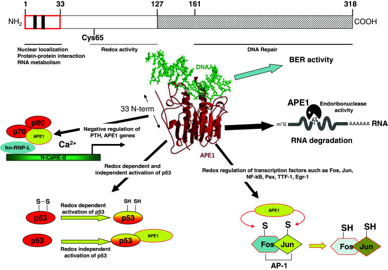
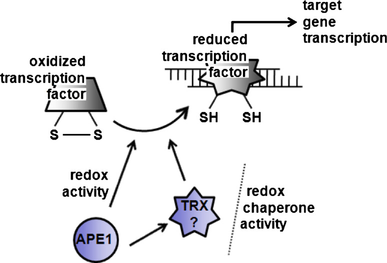
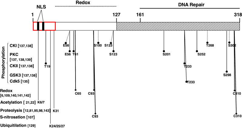
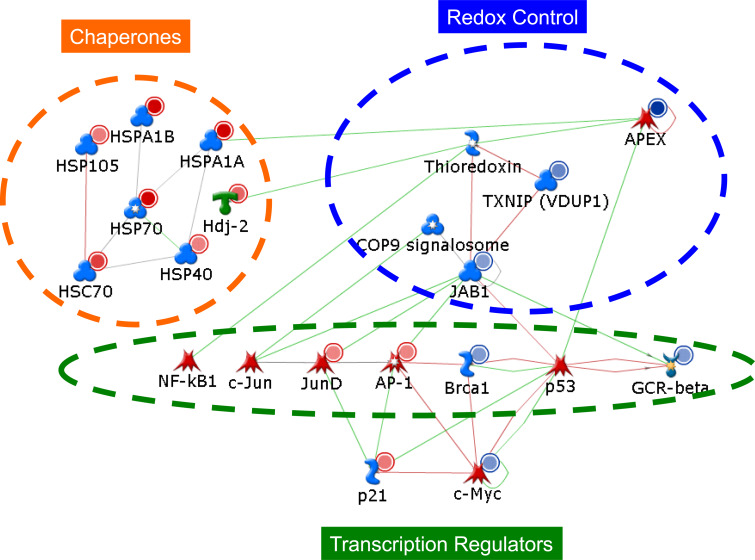
The apurinic endonuclease 1/redox factor-1 (APE1) has a crucial function in DNA repair and in redox signaling in mammals, and recent studies identify it as an excellent target for sensitizing tumor cells to chemotherapy. APE1 is an essential enzyme in the base excision repair pathway of DNA lesions caused by oxidation and alkylation. As importantly, APE1 also functions as a redox agent maintaining transcription factors involved in cancer promotion and progression in an active reduced state. Very recently, a new unsuspected function of APE1 in RNA metabolism was discovered, opening new perspectives for this multifunctional protein. These observations underline the necessity to understand the molecular mechanisms responsible for fine-tuning its different biological functions. This survey intends to give an overview of the multifunctional roles of APE1 and their regulation in the context of considering this protein a promising tool for anticancer therapy.


Figures

Fig. 1
Fig. 1
Schematic organization of human APE1 structure, based on functional studies reported in Refs. [26, 46, 136], with some critical residues. Ribbon representation of the APE1 structure. An α/β-sandwich is formed by the packing of two–six-stranded β-sheets surrounded by α-helices, with strand order β3-β4-β2-β1-β1-β13 and β5-β6-β7-β8-β12-β9 from domains 1 and 2, respectively [14]. The main functions of APE1 are those involved in: (1) BER pathway of DNA lesions; (2) transcriptional repressor through binding to negative calcium responsive elements (N-CaRE); (3) redox-dependent and -independent regulation of different transcription factors activities; (4) RNA metabolism
Fig. 2
Fig. 2
Schematic representation of APE1 redox and redox-chaperone activities. APE1 is responsible for the reduction of oxidized cysteines within DNA-binding domain or regulatory domain of a number of transcription factors. The exact mechanism by which APE1 achieves this thiol reduction is not very well understood. In addition, the state of oxidation of the transcription factor in the nucleus is also poorly characterized (adapted from Ref. [38])
Fig. 3
Fig. 3
Role of APE1 in intracellular ROS production control. APE1 is capable of inhibiting intracellular H2O2 production, through modulation of a Rac1-regulated NADPH oxidase. The molecular mechanism underlying this APE1 activity is not clear; however, a functional interaction between APE1 and Rac1 was demonstrated (adapted from Ref. [48])
Fig. 4
Fig. 4
Schematic representation of APE1 gene promoter region. In the distal promoter region (between −3,000 and −1,000 bp relative to the transcription start site) there are three nCaRE elements; in the proximal promoter region (between −500 and +200 relative to the transcription start site), there are two Sp1 binding elements, an Egr-1 binding site and a CCAAT box. Moreover, several other putative binding sites for other transcription factors are also present
Fig. 5
Fig. 5
Schematic representation of the PTM acceptor sites on APE1 sequence. Although several putative phosphorylation sites have been mapped, it is still unknown which are the real targets of phosphorylation in vivo except for T233 [135]. In contrast, the sites of acetylation, S-nitrosation and ubiquitination of the protein have been precisely mapped on APE1 sequence (adapted from Ref. [2])
Fig. 6
Fig. 6
Different model networks for APE1-mediated signaling as derived by gene expression and proteomic data. Direct interaction network for genes dysregulated upon APE1 knock-down, involved in redox control, stress response and in transcription factors-signaling. Colors of the symbols indicate inhibition (blue) and activation (red). Here, the name APEX is used to refer to APE1. This model network is accessible at this site: https://portal.genego.com/pub/network/n-860173808.html

References

    1. Fung H, Demple B. A vital role for Ape1/Ref1 protein in repairing spontaneous DNA damage in human cells. Mol Cell. 2005;17:463–470. doi: 10.1016/j.molcel.2004.12.029. - DOI - PubMed
    1. Tell G, Damante G, Caldwell D, Kelley MR. The intracellular localization of APE1/Ref-1: more than a passive phenomenon? Antioxid Redox Signal. 2005;7:367–384. doi: 10.1089/ars.2005.7.367. - DOI - PubMed
    1. O’Hara AM, Bhattacharyya A, Bai J, Mifflin RC, Ernst PB, Mitra S, Crowe SE. Tumor necrosis factor (TNF)-alpha-induced IL-8 expression in gastric epithelial cells: role of reactive oxygen species and AP endonuclease-1/redox factor (Ref)-1. Cytokine. 2009;46:359–369. doi: 10.1016/j.cyto.2009.03.010. - DOI - PMC - PubMed
    1. Chung U, Igarashi T, Nishishita T, Iwanari H, Iwamatsu A, Suwa A, Mimori T, Hata K, Ebisu S, Ogata E, Fujita T, Okazaki T. The interaction between Ku antigen and REF1 protein mediates negative gene regulation by extracellular calcium. J Biol Chem. 1996;271:8593–8598. doi: 10.1074/jbc.271.15.8593. - DOI - PubMed
    1. Kuninger DT, Izumi T, Papaconstantinou J, Mitra S. Human AP-endonuclease 1 and hnRNP-L interact with a nCaRE-like repressor element in the AP-endonuclease 1 promoter. Nucleic Acids Res. 2002;30:823–829. doi: 10.1093/nar/30.3.823. - DOI - PMC - PubMed

Publication types

MeSH terms

Substances