Comparative Study
. 2010 Dec;11(4):605-23.
doi: 10.1007/s10162-010-0228-1. Epub 2010 Aug 13.

Divergent aging characteristics in CBA/J and CBA/CaJ mouse cochleae

Affiliations
Comparative Study

Divergent aging characteristics in CBA/J and CBA/CaJ mouse cochleae

Kevin K Ohlemiller et al. J Assoc Res Otolaryngol. 2010 Dec.

Abstract

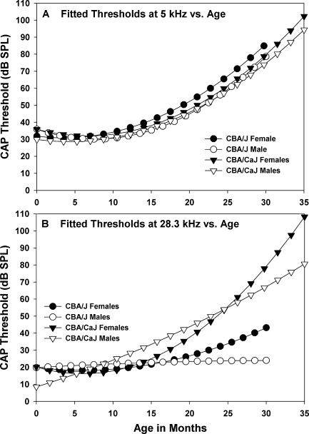
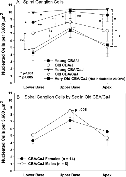
Two inbred mouse strains, CBA/J and CBA/CaJ, have been used nearly interchangeably as 'good hearing' standards for research in hearing and deafness. We recently reported, however, that these two strains diverge after 1 year of age, such that CBA/CaJ mice show more rapid elevation of compound action potential (CAP) thresholds at high frequencies (Ohlemiller, Brain Res. 1277: 70-83, 2009). One contributor is progressive decline in endocochlear potential (EP) that appears only in CBA/CaJ. Here, we explore the cellular bases of threshold and EP disparities in old CBA/J and CBA/CaJ mice. Among the major findings, both strains exhibit a characteristic age (∼18 months in CBA/J and 24 months in CBA/CaJ) when females overtake males in sensitivity decline. Strain differences in progression of hearing loss are not due to greater hair cell loss in CBA/CaJ, but instead appear to reflect greater neuronal loss, plus more pronounced changes in the lateral wall, leading to EP decline. While both male and female CBA/CaJ show these pathologies, they are more pronounced in females. A novel feature that differed sharply by strain was moderate loss of outer sulcus cells (or 'root' cells) in spiral ligament of the upper basal turn in old CBA/CaJ mice, giving rise to deep indentations and void spaces in the ligament. We conclude that CBA/CaJ mice differ both quantitatively and qualitatively from CBA/J in age-related cochlear pathology, and model different types of presbycusis.


Figures

FIG. 1
FIG. 1
AC Mean (−SD) CAP thresholds for CBA/J and CBA/CaJ mice in three different age ranges. Sample size and gender composition of each sample is given in each graph. CBA/CaJ mice show significantly higher thresholds at 20 kHz and above by 17–19 months (B). *Bonferroni multiple comparisons.
FIG. 2
FIG. 2
Scatter plots of CAP threshold versus age in months at 5 kHz (A,B) and 28.3 kHz (C,D) for both CBA/J and CBA/CaJ mice. Left and right columns separate animals by gender. Lines are fitted second-order polynomials. At 28.3 kHz, CBA/CaJ exhibit more hearing loss with age than CBA/J, irrespective of gender.
FIG. 3
FIG. 3
Overlay of fitted polynomial functions from Figure 2. A and B, respectively, superimpose fitted curves for both strains and genders for thresholds at 5 and 28.3 kHz.
FIG. 4
FIG. 4
Mean (+SD) inner and outer hair cell survival for old CBA/J (22–26 months; n = 8) and CBA/CaJ (23–27 months; n = 8) mice. Each group was roughly evenly split by gender. Frequency map is based on Muller et al. (2005). Outer hair cell survival differed significantly by strain and location, with CBA/CaJ mice showing better OHC survival. Horizontal bars above the X axis denote locations where differences were significant by Bonferroni multiple comparisons test.
FIG. 5
FIG. 5
Mean (−SD) spiral ganglion cell density at three cochlear locations by strain and age (A), and for old male and female CBA/CaJ (B). Old CBA/CaJ mice showed significantly greater neuronal loss, particularly in the apex and lower base. Old female CAB/CaJ tended toward greater neuronal loss than males. In B, data from 13 ‘old’ and nine ‘very old’ CBA/CaJ mixed-gender data sets were combined. P values are from Bonferroni multiple comparisons tests following two-way ANOVA.
FIG. 6
FIG. 6
Lower basal turn (mid-hook region) Rosenthal’s canal and organ of Corti in an old female CBA/CaJ mouse showing loss of spiral ganglion cells (SpG). Inset shows expanded organ of Corti, calling attention to the presence of outer hair cells (arrowheads in both images), but the absence of inner hair cells (asterisk) in this animal. SpLim spiral limbus, IP inner pillar, OP outer pillar, D Deiters’ cells.
FIG. 7
FIG. 7
Scatter plots of basal turn EP versus age for CBA/J (top) and CBA/CaJ (bottom). Significant reduction (indicated by significant non-zero regression coefficient) is seen only in CBA/CaJ. Most of the mice with EPs below 95 mV are female.
FIG. 8
FIG. 8
Scatter plots of basal turn EP versus apical turn EP for CBA/J (A) and CBA/CaJ (B) across all ages. Normal EP is ∼10 mV higher in the base than in the apex. Significant EP reduction in CBA/CaJ is associated with reversal of the EP spatial gradient.
FIG. 9
FIG. 9
Distribution of basal turn EPs plotted separately for CBA/J and CBA/CaJ mice during the first 12 months of life (A, B) versus later ages (C, D). Only older CBA/CAJ showed leftward skewing of EPs and dropout of females from EPs >110 mV (arrows). Bins are normalized to sample size by strain and age, so that each bin represents the proportion of animals with an EP in a given range.
FIG. 10
FIG. 10
A Mean(+SD) basal turn EP by strain for animals >12 months old. Only old CBA/CaJ females differed significantly from other groups. B Basal turn EP versus CAP threshold at 28.3 kHz plotted separately for all CBA/CaJ males and females. Significant correlations were found for each. P values in A are from Bonferroni multiple comparisons tests following one-way ANOVA.
FIG. 11
FIG. 11
AF Example lateral wall of the cochlear upper basal turn in six old CBA/CaJ mice with very different EPs. Age, gender, and EP are given in each panel. General features include a mainly normal appearance of the spiral ligament and a large degree of heterogeneity in the stria vascularis. The most glaring pathology of the lateral wall is the apparent loss of outer sulcus cells/root cells in the region of spiral ligament just below spiral prominence (arrows). Asterisk in B highlights large capillaries that appeared most prominent in old female mice (see text). Labels in A denote major landmarks: StV stria vascularis, SM scala media, SpP spiral prominence, I and II types I and II fibrocytes.
FIG. 12
FIG. 12
Mean (−SD) strial thickness versus cochlear location for young and old CBA/J and CBA/CaJ mice. Significant differences were found by strain and age. Both strains showed strial thinning with age, but old CBA/CaJ mice tended toward a thicker stria than old CBA/J. P values are from Bonferroni multiple comparisons tests following two-way ANOVA.
FIG. 13
FIG. 13
A Mean (−SD) number of capillaries per strial profile versus cochlear location for young and old CBA/J and CBA/CaJ. Old CBA/CaJ showed significantly greater capillary loss, yet the magnitude of loss was <20%. B Distribution of strial capillary sizes across all animals, grouped by strain and age. Each symbol represent the proportion of all capillaries falling within a 5 μm diameter range (0–5, 6–10, etc.). Numbers in parentheses are the number of capillaries measured. For young mice of both strains, essentially all capillaries had diameters of 10 μm or less, and appeared uniformly distributed across this range. With age, most animals showed a redistribution of capillary sizes to favor diameters 5 μm or smaller. Old CBA/CaJ females alone (arrow) also featured a notable fraction of capillaries (∼11%) with diameters >10 μm. C Re-plot of data from B normalized using ‘young’ data to show the change in distribution of capillary size with age. Note decrease in proportion of capillaries with diameters of 6–10 μm for all groups, but less pronounced in CBA/CaJ. Arrows highlight qualitatively different redistribution of capillary size in old CBA/CaJ females, including a gain in the fraction of large capillaries. P values in A are from Bonferroni multiple comparisons tests following two-way ANOVA.
FIG. 14
FIG. 14
Mean (+SD) strial marginal cell density at three cochlear locations by strain and age. Young CBA/J mice tended toward more marginal cells than young CBA/CaJ, and sustained no significant loss with age. Old CBA/CaJ showed significant loss of marginal cells, particularly in the lower cochlear base, with no clear effect of gender (not shown). P values are from Bonferroni multiple comparisons tests following two-way ANOVA.
FIG. 15
FIG. 15
Scatter plots of three anatomic metrics in the upper basal turn (marginal cell density, OSC abnormality, strial capillary density) versus basal turn EP for CBA/CaJ (AC) and CBA/J (df). Only marginal cell density in CBA/CaJ was significantly correlated with EP.
FIG. 16
FIG. 16
Mean (−SD) spiral ligament thickness versus cochlear location for young and old CBA/J and CBA/CaJ mice. Significant differences were found by strain, so that CBA/CaJ mice showed a thinner ligament, particularly in the basal half of the cochlea. P values are from Bonferroni multiple comparisons tests following two-way ANOVA.
FIG. 17
FIG. 17
Example lateral wall of the cochlear upper basal turn in an old CBA/CaJ female (A same animal as in Fig. 11A) and an old CBA/J female (B). Age and EP in each mouse are shown. The two images have been aligned to emphasize difference in survival of outer sulcus cells/root cells in the region just below spiral prominence (compare locations at large arrows). Potentially related was a difference in the extent of coverage of OSCs at this location by spiral prominence epithelial cells. In CBA/J mice, the cells of spiral prominence (SpP) more often contact Claudius cells (Cc) of the organ of Corti. Small arrowheads in each panel denote the apparent end of Claudius cell processes, leaving more OSCs exposed to endolymph in the CBA/CaJ.
FIG. 18
FIG. 18
A Mean(-SD) outer sulcus cell density in the upper and lower basal turn of young and old CBA/J and CBA/CaJ mice. CBA/CaJ mice show fewer of these cells, irrespective of age. B Incidence of abnormal OSC/root cell profiles by strain and age. Uniquely in old CBA/CaJ mice, over 60% of sections exhibited indentations or voids in spiral ligament suggestive of OSC/root cell loss. P values are in A from Bonferroni multiple comparisons tests following two-way ANOVA.

Similar articles

Cited by

References

    1. Adams JC, Schulte BA. Histopathologic observations of the aging gerbil cochlea. Hear Res. 1997;104:101–111. doi: 10.1016/S0378-5955(96)00184-0. - DOI - PubMed
    1. Bohne BA, Gruner MM, Harding GW. Morphological correlates of aging in the chinchilla cochlea. Hear Res. 1990;48:79–91. doi: 10.1016/0378-5955(90)90200-9. - DOI - PubMed
    1. Bult CJ, Eppig JT, Kadin JA, Richardson JE, Blake JA. The mouse genome database (MGD): mouse biology and model systems. Nucleic Acids Res. 2008;36:D724–D728. doi: 10.1093/nar/gkm961. - DOI - PMC - PubMed
    1. Charitidi K, Meltser I, Tahera Y, Canlon B. Functional responses of estrogen receptors in the male and female auditory system. Hear Res. 2009;252:71–78. doi: 10.1016/j.heares.2008.12.009. - DOI - PubMed
    1. Cohen GM, Park JC, Grasso JS. Comparison of demyelination and neural degeneration in spiral and Scarpa's ganglion of C57BL/6 mice. J Electron Microsc Tech. 1990;15:165–172. doi: 10.1002/jemt.1060150208. - DOI - PubMed

Publication types