Review
. 2011:62:431-45.
doi: 10.1146/annurev-med-052209-100430.

Stress- and allostasis-induced brain plasticity

Affiliations
Review

Stress- and allostasis-induced brain plasticity

Bruce S McEwen et al. Annu Rev Med. 2011.

Abstract

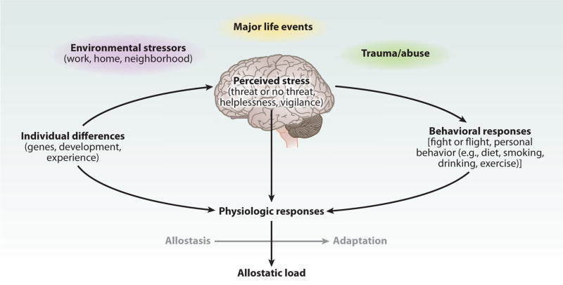
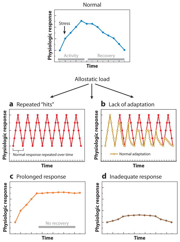
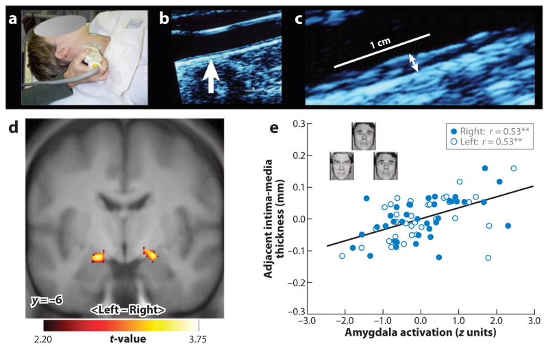
The brain is the key organ of stress processes. It determines what individuals will experience as stressful, it orchestrates how individuals will cope with stressful experiences, and it changes both functionally and structurally as a result of stressful experiences. Within the brain, a distributed, dynamic, and plastic neural circuitry coordinates, monitors, and calibrates behavioral and physiological stress response systems to meet the demands imposed by particular stressors. These allodynamic processes can be adaptive in the short term (allostasis) and maladaptive in the long term (allostatic load). Critically, these processes involve bidirectional signaling between the brain and body. Consequently, allostasis and allostatic load can jointly affect vulnerability to brain-dependent and stress-related mental and physical health conditions. This review focuses on the role of brain plasticity in adaptation to, and pathophysiology resulting from, stressful experiences. It also considers interventions to prevent and treat chronic and prevalent health conditions via allodynamic brain mechanisms.


Figures

Figure 1
Figure 1
Central role of the brain in allostasis and the behavioral and physiological response to stressors. Redrawn from Reference 5 with permission.
Figure 2
Figure 2
Types of allostasis and allostatic load. (a) Repeated “hits” from multiple stressors. Individuals who are repeatedly exposed to stressors over their life course and who also experience large surges in blood pressure and cardiovascular activity, which depend on the engagement of the hypothalamic-pituitary-adrenal (HPA) and autonomic axes, are more likely to show premature hypertension and atherosclerotic heart disease. (b) Lack of adaptation. A failure to habituate to a repetition of the same stressor results in a persistent elevation of mediators such as cortisol. This was first described in individuals who failed to habituate in their cortisol response to a public-speaking stressor (5, 71). (c) Prolonged response due to delayed shutdown. Adaptive autonomic and neuroendocrine responses are slow to terminate; e.g., blood-pressure elevations are sustained during repetitive, time-pressured work. (d) Inadequate response leads to compensatory hyperactivity of other mediators. For example, autoimmunity and inflammation can be associated with inadequate endogenous glucocorticoid responses, as in the Lewis rat and possibly also in chronic fatigue syndrome and fibromyalgia.
Figure 3
Figure 3
In a functional neuroimaging study, a measure of preclinical atherosclerosis was related to activation of the amygdala in response to threatening facial expressions. Carotid intima-media thickness (IMT) was assessed by B-mode ultrasonography. (a) A trained vascular technologist imaged the carotid arteries. (b) An image of the carotid artery with the common carotid segment visible at right and the beginning of the carotid bulb visible at left; arrow indicates a point along the far wall of the common carotid artery, which is shown at higher magnification in panel c. (c) Magnified point of the far wall of the common carotid artery illustrating the lumen-intima and media-adventitia interfaces. IMT was measured by averaging the IMT (the distance between two lines tracking the lumen-intima and media-adventitia interfaces, not illustrated here) in 1-mm increments along the distal 1 cm of the far wall of the common carotid artery, the far wall of the carotid bulb, and the first centimeter of the internal carotid artery. As assessed by this ultrasound procedure, carotid IMT covaried positively with amygdala reactivity to angry and fearful facial expressions in an adult sample of otherwise healthy humans. (d) Statistical parametric maps from a regression analysis identifying regions of the left and right amygdala where carotid IMT covaried with amygdala reactivity after controlling for age, sex, resting systolic blood pressure, and family income. (e) Adjusted IMT values are shown as a function of amygdala activation values of the left (open circles) and right (closed circles) amygdala areas profiled in panel d. Insets in panel e illustrate sample trials from the facial-expression protocol designed to elicit amygdala reactivity. **p < 0.001.

References

    1. Cohen S, Janicki-Deverts D, Miller GE. Psychological stress and disease. JAMA. 2007;298:1685–88. - PubMed
    1. McEwen BS. Physiology and neurobiology of stress and adaptation: central role of the brain. Physiol Rev. 2007;87:873–904. - PubMed
    1. Lazarus RS, Folkman S, editors. Stress, Appraisal and Coping. New York: Springer-Verlag; 1984.
    1. Knudsen EI, Heckman JJ, Cameron JL, et al. Economic, neurobiological, and behavioral perspectives on building America's future workforce. Proc Natl Acad Sci USA. 2006;103:10155–62. - PMC - PubMed
    1. McEwen BS. Protective and damaging effects of stress mediators. N Engl J Med. 1998;338:171–79. - PubMed

Publication types